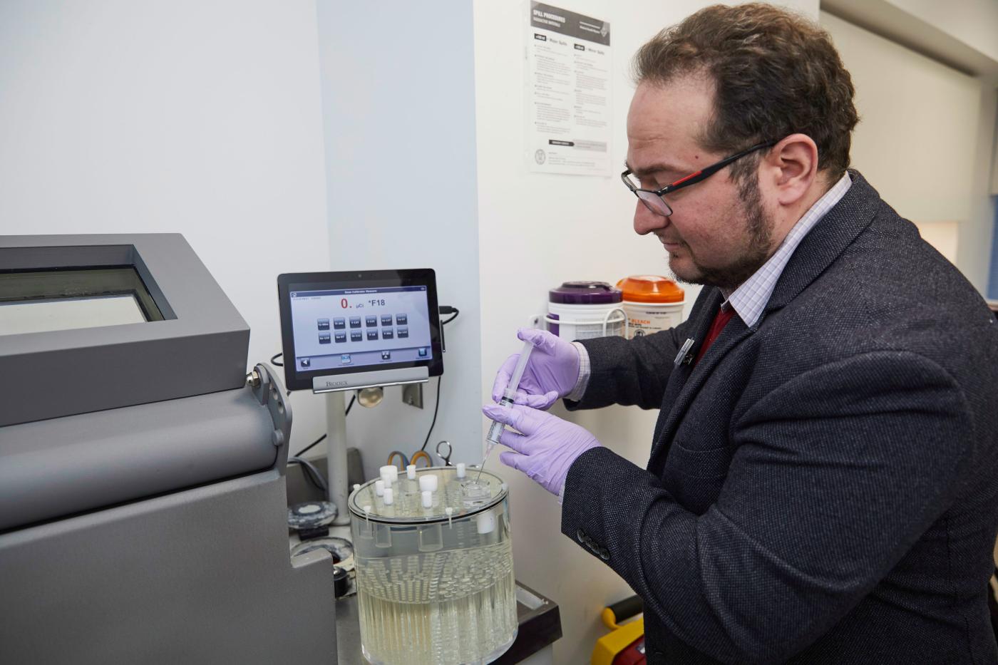
Dr. Nikolaos (Nicolas) A. Karakatsanis has been an assistant professor of biomedical engineering and a nuclear medicine physicist in the radiology department at Weill Cornell Medicine (WCM) since 2017. He is also an adjunct professor in the Biomedical Engineering and Imaging Institute (BMEII) of the Icahn School of Medicine at Mount Sinai Hospital. His research interests include quantitative whole-body dynamic positron emission tomography (PET) to enable the routine clinical adoption of multi-parametric PET imaging for enhanced diagnostic and therapy response PET assessments in oncology, cardiology and neurology. He has developed dynamic PET-driven quantitative methods to improve attenuation correction and dual-radiotracer decoupling methods in simultaneous PET/magnetic resonance (MR) imaging largely for cardiovascular disease evaluations. He is developing cost-effective long axial field-of-view (AFOV) sparse PET detection systems to facilitate the widespread clinical adoption of the quantitative virtues of total-body dynamic PET imaging.
Dr. Karakatsanis has co-authored more than 50 peer-reviewed original articles and more than 80 conference records in national and international scientific meetings. A member of the Society of Nuclear Medicine and Molecular Imaging (SNMMI), he has served as intern (2017-2019) and secretary officer (2019-2021) in the Physics, Data Sciences and Instrumentation Council (PIDSC). He is the founder and administrator of NMMItools.org, a web-resource page developed during his PIDSC internship to host an up-to-date list of validated commercial and open-source software tools to support state-of-the-art research in nuclear medicine and molecular imaging. Dr. Karakatsanis is a senior member of the Institute of Electrical and Electronic Engineers (IEEE) and has served as member-at-large (2017-2020) and as the secretary officer (since 2021) of the IEEE Nuclear Medical Imaging Sciences Council (NMISC). Dr. Karakatsanis is certified by the American Board of Science in Nuclear Medicine (ABSNM) in nuclear medicine physics and instrumentation. He is an associate editor with Frontiers of Medicine and Clinical Imaging and serves as guest editor with Medical Physics.
Associated Lab Members

Dr. Nikolaos (Nicolas) A. Karakatsanis has been an assistant professor of biomedical engineering and a nuclear medicine physicist in the radiology department at Weill Cornell Medicine since 2017. He previously held research scientist positions in the Icahn School of Medicine at Mount Sinai Hospital (2015-2017), University Hospitals of Geneva, Switzerland (2014-2015) and Johns Hopkins University Hospital (2011-2013). Dr. Karakatsanis received his masters’ degree, and his Ph.D., in Electrical and Computer Engineering in 2005 and 2010 respectively from the National Technical University of Athens, Greece.
In terms of PET data acquisition protocols, the lab is designing clinically adoptable dynamic whole-body human PET scan protocols customized for the short AFOV human PET scanners that are widely available today. The aim is to utilize those imaging protocols to translate the quantitative virtues of truly multi-parametric, beyond static SUV PET imaging to oncology, cardiology, and multi-organ (e.g., brain-heart axis) studies. As a pioneer in the development of the first dynamic whole-body 18 fluorodeoxyglycose (FDG) PET/CT human scan protocols a few years ago, the lab now collaborates closely with nuclear medicine and radiology physicians to expand and customize those PET protocols for other important radiotracers such as 68 gallium (Ga)-prostate specific membrane antigen (PSMA) and 68Ga-DOTATATE to benefit diagnosis and treatment response assessment for metastatic castrate-resistant prostate cancer (mCRPC), neuroendocrine tumors (NETs), and cardiovascular diseases. The lab is investigating novel dual-radiotracer PET/MR acquisition protocols, with 18F-sodium fluoride (NaF) being one of the two administered radiotracers acting as an agent of unique dynamics to drive bone segmentation and aid in the synthesis of more complete tissue attenuation maps. The result: more accurate attenuation correction of the detected PET signal in PET/MR studies where CT modality, the gold standard for PET attenuation correction, is missing.
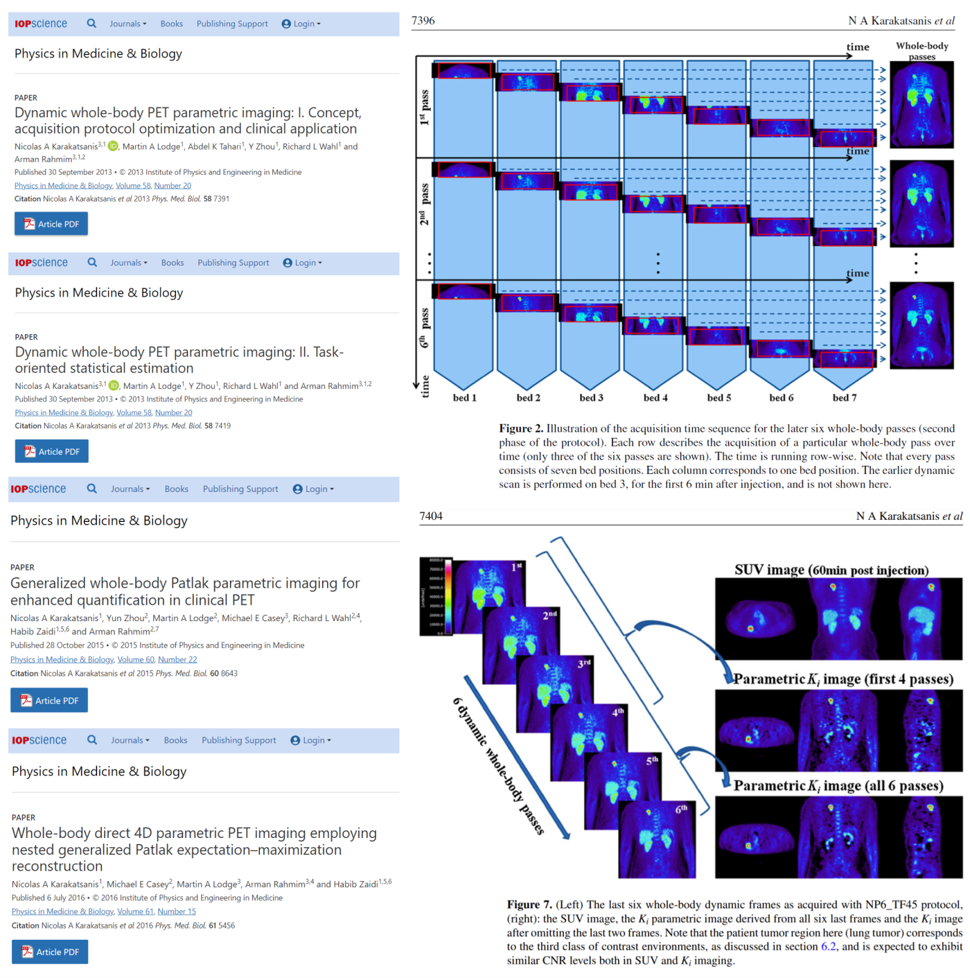
Regarding PET image reconstruction, the lab introduced novel direct 4D PET image reconstruction algorithms tailored for multi-bed or continuous bed motion acquisitions to support multi-parametric whole-body PET imaging even with the limited AFOV clinical PET scanners widely accessible now. In those algorithms, the lab incorporated robust graphical analysis methods enabling direct imaging of multiple clinically relevant macro-kinetic parameters of the administered radiotracers with high precision and accuracy despite the high noise levels typically present in the dynamic whole-body raw PET data. Aside from their robustness, the utilized graphical analysis methods can significantly shorten the total scan period required to capture the macro-dynamics of the administered radiotracer. The lab aims to match the scan periods post-injection (p.i.) that are typically used for routine standard-of-care static PET scan protocols, e.g., 60-80 min p.i. in the case of whole-body 18F-FDG PET exams, without compromising for kinetic macro-parameters accuracy and precision. The lab is collecting independent measurements of specific radiotracers’ concentration in arterial blood plasma, known as input function, from respective populations of past dynamic PET exams to build population-based models for each specific radiotracer. These population-based models let the lab acquire prospectively only a small portion of the input function at later and shorter time windows post-administration, matching the time window employed for typical state-of-the-art static PET scans, allowing for inference of the missing earlier section from the population-based model.
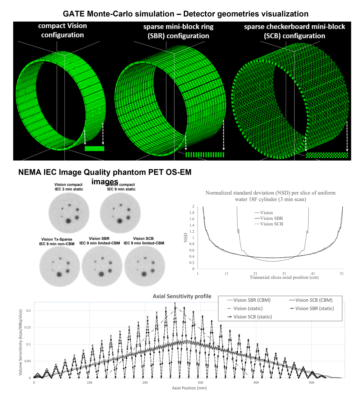
The lab also investigates novel methods of data normalization and machine-learning (ML) aided image reconstruction customized for PET data acquired with cost-effective sparse PET detector geometries with double the AFOV of the respective compact PET detector geometries. To attain high image quality for reconstructed sparse PET data, similar to that attained with compact PET scanners, the lab employed a customized component-based normalization method accounting for the sparse sampling effects due to gaps presence. The lab developed a novel limited continuous bed motion (limited-CBM) PET acquisition mode for the sparse geometries to smooth the expected large variance in axial sensitivity observed between the large gaps and the physical detector blocks. The lab is also investigating, with other Cornell artificial intelligence experts, ML approaches based on U-net deep convolutional neural networks (CNNs) to recover losses in image quality and mitigate elevated noise levels due to sparse sampling reconstructing and analyzing sparse PET data. The lab sees this as very important as it can allow the development of cost-effective clinical PET scanners of adaptable AFOVs supporting, under a single universal scanner architecture, multiple customized configurations optimized for the unique demands of each research or clinical PET study. Future generation clinical PET scanners may adapt their configuration optimally to offer either highly sensitive scans over shorter AFOVs, or moderately sensitive scans over longer AFOVs, depending on the targeted imaging task of each study.
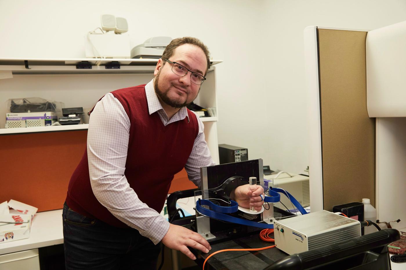
The lab is developing a realistic Monte-Carlo (MC) simulation of a dedicated mini-positron emission tomography (PET) cylindrical scanner designed for the direct non-invasive tomographic measurement of the dynamic activity concentration of a PET...
The lab has recently introduced at Weill Cornell Medicine (WCM) the concept of a cost-effective positron emission tomography (PET) detector geometry with adaptable axial field of view (AFOV) via a published series of realistic Monte Carlo simulation...
Prostate cancer is one of the leading causes of cancer death among men in the U.S. and worldwide. The Prostate-Specific Membrane Antigen (PSMA) is a transmembrane protein expressed in all types of prostatic tissue and is broadly recognized as a useful...