One of modern biomedical science's greatest challenges is the mapping of the human brain to understand functionality and behavior. UHF MRI (using magnetic field strengths of 7T and above) offers promise of a crucial improvement over 3T in spatial resolution and sensitivity for deciphering subtle features that are <1mm in size and could allow mapping of intricate detail such as intra-cortical or small subcortical network hubs.
Substantial technological hurdles still hinder UHF field strengths before performance increases toward higher sensitivity and sub-mm resolution are fully realized. The Winkler Lab focuses on an interdisciplinary approach at the intersection of engineering and medicine to overcome key hardware and technological critical barriers that are holding back UHF MRI and its clinical applicability.
Associated Lab Members
Professor Simone Angela Winkler graduated from the J. Kepler University of Linz, Austria, having majored in mechatronics with distinction and in less than minimum time. She pursued her graduate studies in electrical engineering at the École Polytechnique Montréal, Canada, where she specialized in radiofrequency (RF)/microwave engineering, funded by two fellowships (DOC fellowship/Austrian Academy of Science; first rank in the competition for a PhD fellowship from the FQRNT Québec). For her research during her MS.c. and Ph.D. degrees, she received many scientific awards and scholarships. During her postdoctoral work at McGill University, she developed a microwave near-field imaging system for breast cancer detection. She committed to a postdoctoral fellow position at Stanford University in ultra-high-field magnetic resonance imaging engineering (funded by a National Sciences and Engineering Research Council of Canada (NSERC) research fellowship from 2012-2014).
Elizaveta Motovilova received her B.Sc. and M.Sc. with a major in applied mathematics and physics from the Moscow Institute of Physics and Technology in 2012 and 2014, respectively. She received her Ph.D. from the Singapore University of Technology and Design (SUTD) in 2019. During her studies at SUTD, she was awarded the Institute of Electrical and Electronics Engineers (IEEE) MTT-S Microwave Engineering for Medical Applications Fellowship for research on the sensitivity improvement of radiofrequency (RF) coils for magnetic resonance imaging (MRI). From 2019 to 2020, she was a postdoctoral research fellow at SUTD, where she continued her work on MRI radiofrequency (RF) coils with a focus on frequency tuning mechanisms. Her main research interests include design and development of MRI RF coils, metamaterials and resonators for RF coil sensitivity improvement, ultra-high field MRI engineering and safety.
Nazish Murad, Ph.D., received her doctorate in biomedical engineering from National Central University, Taiwan. A research enthusiast with a passion for unraveling machine learning and its biomedical imaging applications, Dr. Murad has been honored with prestigious scholarships and awards, including the Ph.D. fellowship from the National Central University and CTCI Research Scholarship. Dr. Murad specializes in optical image reconstruction—signal-to-image biomedical imaging—and has applied myriad machine-learning techniques to tackle medical challenges. Passionate about innovation and mentorship, Dr. Murad thrives on pushing the boundaries of knowledge and making impactful contributions to the scientific community.
Rigoberto Vazquez Jr. earned his B.Sc. in Mechanical Engineering from California State University in 2019 and his M.Sc. in Nuclear Engineering & Radiological Science—with emphasis in Materials and Medical Physics—from the University of Michigan in 2021. Vazquez is now pursuing his Ph.D. in the biomedical engineering program administered by the Weill Cornell Graduate School of Medical Sciences and Cornell University Graduate School. His current research interest focuses on the design and development of MRI RF coils for nonconventional anatomical regions using conductive materials and a variety of 3D-printing techniques.
The Winkler Lab investigates novel hardware and technology methods in UHF MRI, promising crucial improvement in spatial resolution and sensitivity for deciphering subtle features that are <1mm in size.
The lab fosters collaborations with institutions worldwide, particularly General Electric (GE) Healthcare, Stanford University, and inGenuyX engineering solutions.
The lab welcomes a diverse set of talents. It fosters an interdisciplinary approach at the intersection of the engineering and medical fields in its attempts to translate fundamental scientific discoveries into clinical innovations.
Follow this LINK for a set of slides that gives an overview of past research topics on UHF MRI at Stanford University.
A great challenge of modern biomedical science is the mapping of the human brain to understand underlying functionality and behavior. The National Institutes of Health (NIH)-funded Human Connectome Project (HCP) is a large-scale,...
The Winkler Lab has a dedicated radiofrequency (RF) team with a top-notch RF lab to design, demonstrate and test high field and UHF receive and transmit coils. Our team is devoted to cutting-edge research studies t...
Magnetic resonance imaging (MRI) relies on a dense array of radiofrequency (RF) coils to obtain functional and anatomical information inside the body. Tightly fitting coil arrays boost the signal to noise ratio (SNR) and imaging speed. Unfortunately, most commercial RF coils are rigid,...
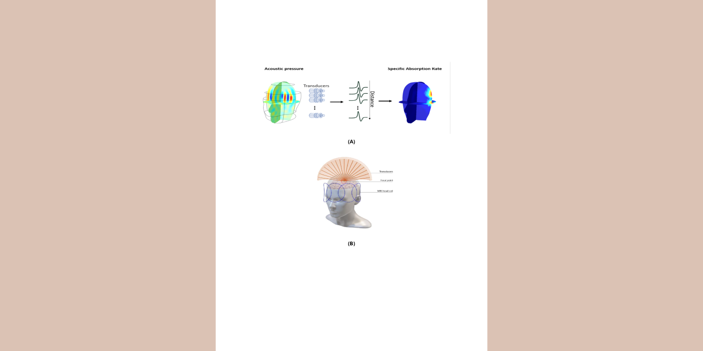
Magnetic resonance (MR)-guided focused ultrasound (MRgFUS) is a non-invasive therapeutic modality for neurodegenerative diseases that allows real-time imaging of targeted regions. However, MR image quality is poor and severely limits the technology due to the use of the body coil for...
A crucial safety concern for ultra-high field (UHF) magnetic resonance imaging (MRI) is the significant radiofrequency (RF) power deposition in the body in the form of local specific absorption rate (SAR) hotspots, leading to dangerous tissue heating/damage. This work is a proof-of-concept demonstration of artificial intelligence (AI)...
The rapid and successful advancement in ultra-high field (UHF) magnetic resonance (MR) scanners (7 tesla (T) or higher) has led to improvement in the spatial and temporal resolution and the signal-to-noise ratio (SNR) per...