Associated Lab Members

Douglas Ballon received his Ph.D. from Rutgers University in 1985 in experimental nuclear physics. He did postdoctoral work in medical physics at Memorial Sloan Kettering Cancer Center from 1985-1988, and subsequently joined the faculty there, developing magnetic resonance imaging (MRI) techniques for applications in oncology. In 2001, he became Founding Director of the Citigroup Biomedical Imaging Center (CBIC) at Weill Cornell Medical College, a comprehensive MRI, positron emission tomography (PET), single photon emission computed tomography (SPECT), CT, ultrasound, optical imaging, and cyclotron facility that supports nearly 100 investigators from 15 academic institutions. Over the last 20 years he has held a leadership role in the development and management of imaging technologies. He has more than 30 years of experience in the development of imaging biomarkers for the detection, characterization, and therapeutic monitoring of disease.

Elizaveta Motovilova received her B.Sc. and M.Sc. with a major in applied mathematics and physics from the Moscow Institute of Physics and Technology in 2012 and 2014, respectively. She received her Ph.D. from the Singapore University of Technology and Design (SUTD) in 2019. During her studies at SUTD, she was awarded the Institute of Electrical and Electronics Engineers (IEEE) MTT-S Microwave Engineering for Medical Applications Fellowship for research on the sensitivity improvement of radiofrequency (RF) coils for magnetic resonance imaging (MRI). From 2019 to 2020, she was a postdoctoral research fellow at SUTD, where she continued her work on MRI radiofrequency (RF) coils with a focus on frequency tuning mechanisms. Her main research interests include design and development of MRI RF coils, metamaterials and resonators for RF coil sensitivity improvement, ultra-high field MRI engineering and safety.

Yun Shang, Ph.D., received his doctorate, M.P.H., and M.Sc. from Columbia University; another M.Sc. from East China Normal University; and his B.E. from Shanghai University of Science and Technology.
Rigoberto Vazquez Jr. earned his B.Sc. in Mechanical Engineering from California State University in 2019 and his M.Sc. in Nuclear Engineering & Radiological Science—with emphasis in Materials and Medical Physics—from the University of Michigan in 2021. Vazquez is now pursuing his Ph.D. in the biomedical engineering program administered by the Weill Cornell Graduate School of Medical Sciences and Cornell University Graduate School. His current research interest focuses on the design and development of MRI RF coils for nonconventional anatomical regions using conductive materials and a variety of 3D-printing techniques.
Imaging techniques for applications in genetic medicine
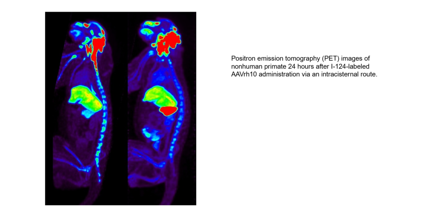
Development of quantitative imaging biomarkers for applications in genetic medicine; in particular, for studying the organ tropism of a range of wildtype and engineered viral vectors used in gene therapy, and for measuring whole-body immune response to vector administration.
Award or Grant: National Institutes of Health (NIH), R01 EB027918
The goal of this project is to develop noninvasive, safe, temporal monitoring of adeno-associated viral vector (AAV) biodistribution following in vivo administration that can be ultimately used in humans. The lab’s strategy is to...
Award or Grant: U54NS065768, BioMarin Pharmaceutical, 190-201, 190-202, 190-203
Ceroid lipofuscionosis type 2 (CLN2) disease is a rare, rapidly progressing lysosomal storage disease with severe neurological complications including widespread neurodegeneration. The disease’s rarity, as well...
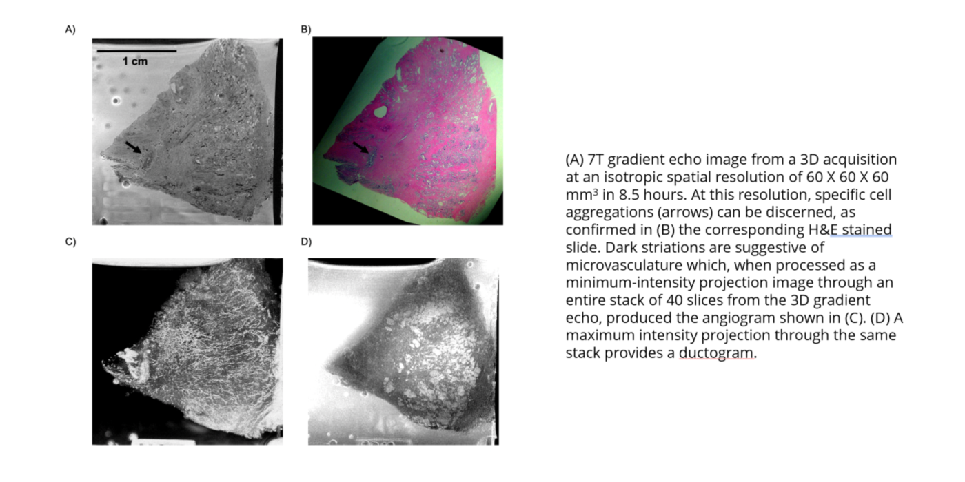
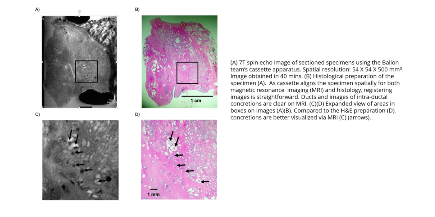
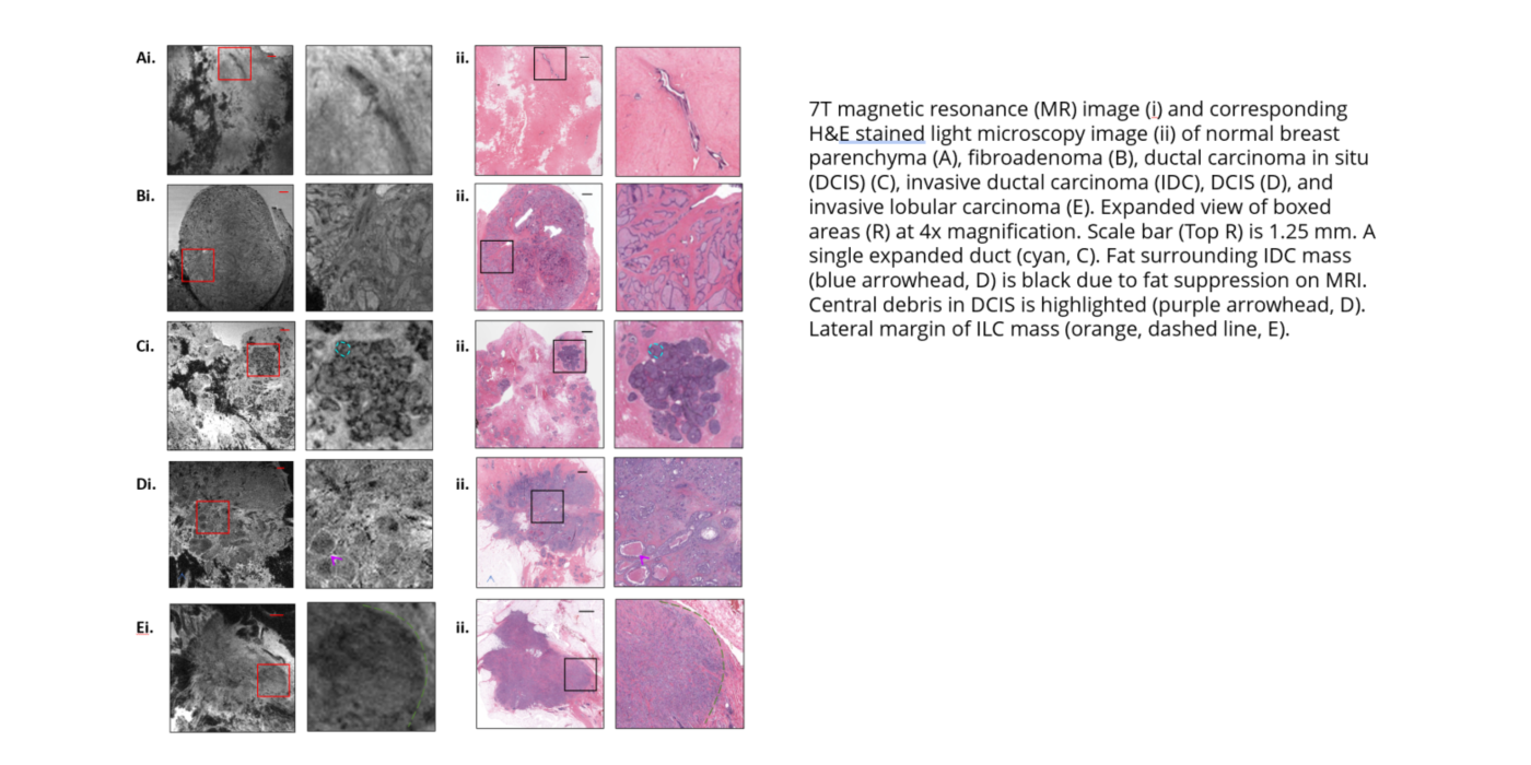
Award or Grant: Radiological Society of North America (RSNA) Research and Education Foundation Roentgen Resident/Fellow Research Award
Residents and fellows rotating through the Ballon lab have developed methods for imaging specimens using magnetic resonance imaging (MRI) at high spatial...
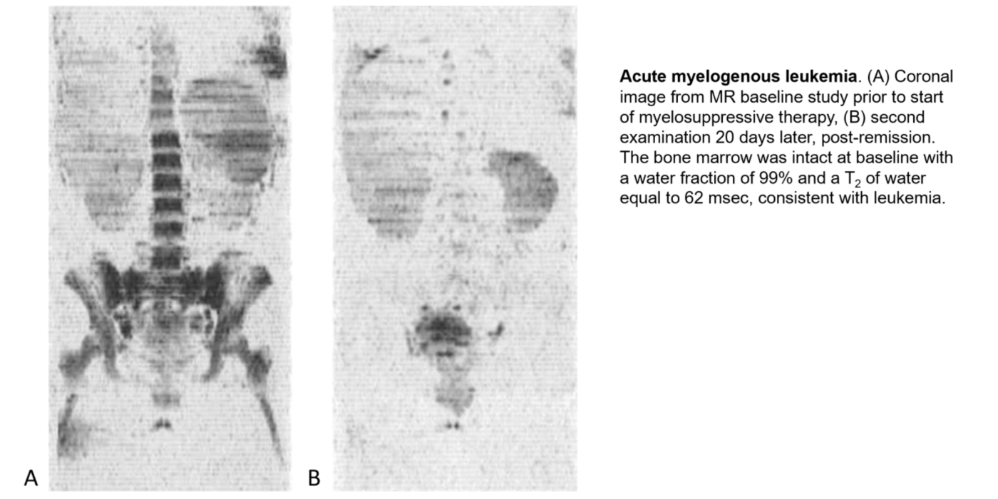
Award or Grant: National Institutes of Health (NIH), R01EB002070
The Ballon lab has developed new methods for whole-body imaging of metastatic disease in oncology. The lab introduced a rapid, whole-body magnetic resonance imaging (MRI) technique for leukemia and metastatic cancers including prostate and breast...
Award or Grant: National Institutes of Health (NIH), S10OD030447
The lab’s research efforts using iodine (I)-124 positron emission tomography (PET) to image viral vectors require a cyclotron for production of the radioisotope. The cyclotron, in turn, requires special targetry to produce iodine (I)-124. The research...