Prostate cancer is one of the leading causes of cancer death among men in the U.S. and worldwide. The Prostate-Specific Membrane Antigen (PSMA) is a transmembrane protein expressed in all types of prostatic tissue and is broadly recognized as a useful diagnostic and therapeutic agent for prostate cancer. Static whole-body (WB) positron emission tomography/computed tomography (PET/CT), using the 68 gallium (Ga)-labelled PSMA-11 (68Ga-PSMA) radiotracer, has been recently adopted in clinics for diagnosing metastatic castrate-resistant prostate cancer (mCRPC) and assessing its response to PSMA-targeted radionuclide treatment (PSMA-TRT). However, the standardized uptake value (SUV) metric of static PET images depends on several factors varying across scans, such as the blood plasma tracer concentration (input function), which are not considered by SUV, thus posing limitations in accurately quantifying prostate activity and treatment response.
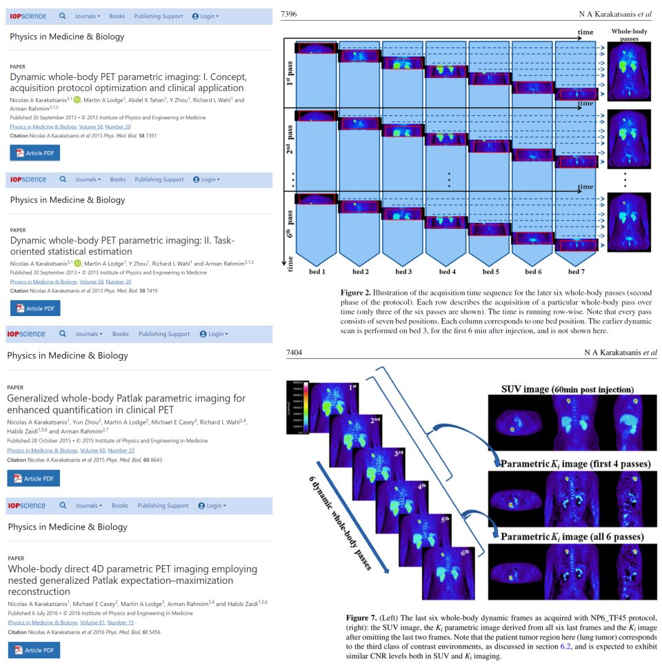
Parametric imaging from 68Ga-PSMA PET/CT dynamic scans may overcome these limitations by accounting for the input function and enabling imaging of unique physiological 68Ga-PSMA parameters beyond the SUV. However, due to the limited axial field-of-view (AFOV) of most clinical PET/CT scanners of today, dynamic 68Ga-PSMA PET protocols are confined to single-bed AFOVs with limited body coverage. Although total body PET scanners may address this limitation, their multi-fold high cost prevents their clinical adoption. The recently introduced dynamic WB 18F-fluorodeoxyglucose (FDG) PET/CT protocols enable WB Patlak imaging of the net uptake rate constant (Ki) from low-cost clinical PET scanners with limited AFOVs. But associated long scan times (>60 min) limit their clinical adoption.
The Karakatsanis Lab is developing a novel dynamic WB 68Ga-PSMA PET/CT framework supporting combined SUV and direct-4D Patlak Ki WB imaging within standard-of-care (SOC) scan time windows post-injection (p.i.). The WB SUV images will be estimated by summing up the 68Ga-PSMA data of all passes, while the WB Patlak Ki images will be reconstructed from the same data using direct-4D Patlak image reconstruction and a population-based input function (pIF) model. The pIF model will be built from a population of image-derived input function data acquired non-invasively. Our methods will be validated using both experimental phantom and clinical data. We hypothesize that multi-parametric SUV/Ki WB 68Ga-PSMA PET/CT within SOC time windows is clinically adoptable and will enhance mCRPC lesion detectability and correlation of PSMA-TRT response assessments with biopsy-confirmed PSA levels. This method can introduce a paradigm shift in the clinic for metastatic castration resistant prostate cancer (mCRPC) diagnosis and PSMA-TRT response utilizing limited AFOV clinical PET/CT scanners.


