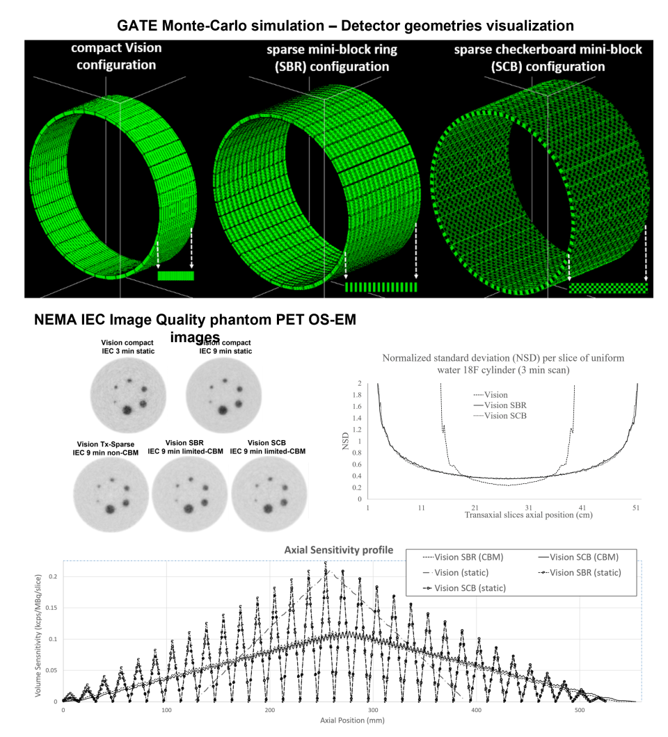
The lab has recently introduced at Weill Cornell Medicine (WCM) the concept of a cost-effective positron emission tomography (PET) detector geometry with adaptable axial field of view (AFOV) via a published series of realistic Monte Carlo simulation studies. The proposed PET systems employ a robust gantry motion mechanism to repeatedly adapt the degree of sparsity between detector blocks to routinely transition between a conventional compact PET detector geometry of a given AFOV length and a sparse PET geometry with the same number of detectors but nearly double its compact AFOV.
During the assessment of its National Electrical Manufacturers Association (NEMA) performance characteristics, a large axial variance between the detectors and the interleaved gaps between them was observed causing image artifacts and elevated statistical noise in the final reconstructed PET images. Currently, the lab is evaluating alternative sparse PET detector configurations of double the current generation scanners axial FOV by interleaving uniforms gaps either every-other-block ring or by forming a checkerboard pattern against the default compact configuration of modern clinical PET scanners. The lab is also developing customized limited continuous bed motion (limited-CBM) acquisition methods coupled with component-based 3D data normalization methods accounting for the presence of gaps to eliminate the large variance in spatial sampling rate and sensitivity. The aim: double the AFOV of existing limited AFOV PET scanners without increasing total detector numbers while delivering high-quality PET images with clinically acceptable lesion detectability performance very similar to that attained with conventional compact PET scanner geometries.
However, the above achievement may still not address the problem of elevated statistical noise observed with sparse PET detector configurations due to the lower sampling density of the central axial FOV region. Therefore, the lab is also working on the development of customized machine learning methods using deep convolutional neural networks to recover within or post reconstruction a higher signal-to-noise ratio in the sparse PET images similar to that attained with compact PET geometries without compromising PET signal quantitation.


