Dr. Mamou’s lab is focused on the development and translation of ultrasound technology to basic science, small-animal, and clinical applications. Dr. Mamou has more than 15 years of National Institutes of Health (NIH)-funded research experience in a variety of ultrasound imaging and therapy topics. Since 2022, he has been at Weill Cornell Medicine (WCM), where he and Dr. Jeffrey Ketterling founded the Biomedical Ultrasound Research Laboratory (BURL).
Dr. Mamou has been involved in the development of quantitative ultrasound (QUS) methods for ultrasound-based tissue characterization. He has advanced the field with new methods and successfully applied QUS method to numerous organs (i.e., lymph nodes, thyroid, prostate, eye, lung, etc.)
Dr. Mamou is also a high-frequency ultrasound (i.e., > 25 MHz) expert who has developed several new methods to improve image quality and penetration depth. His research also includes signal- and image-processing and QUS methods applied to high-frequency ultrasound data.
In the past 10 years, Dr. Mamou has been very involved in the field of quantitative acoustic microscopy which uses ultra-high ultrasound frequencies (i.e., > 250 MHz) to form 2D maps of acoustic properties of thin sections of soft tissues. These maps provide unique information about tissues at microscopic scales and can better our understanding of numerous diseases where mechanical properties of tissues are affected.
Associated Lab Members

Dr. Mamou is a professor of electrical engineering in radiology at Weill Cornell Medicine. Jonathan earned a B.S. in electrical engineering from the Ecole Nationale Superieure des Telecommunications in Paris, France, in 2000. He earned a Ph.D. in electrical engineering in 2005 from the University of Illinois at Urbana-Champaign. His fields of interest include theoretical aspects of ultrasonic scattering, ultrasonic medical imaging, ultrasound contrast agents, and biomedical image processing. Recent projects focus on quantitative ultrasound methods for ultrasound tissue characterization in oncology and ophthalmology, and ultra-high frequencies (>250 MHz) for quantitative acoustic microscopy, among others.

Dr. Jeff Ketterling is a professor of biomedical engineering in radiology at Weill Cornell Medicine. Jeff received his B.S. in electrical engineering from the University of Washington in 1994, and his Ph.D. in mechanical engineering from Yale University in 1999. Jeff’s research focuses on the development and translation of ultrasound technology to basic science, and small-animal and clinical applications, particularly in the realm of high-frequency ultrasound. Recent projects include high-speed plane-wave ultrasound imaging for intracardiac flow patterns in mouse adult mice, blood flow in the front and back of the human eye, and activation of acoustic nanodrops for imaging microcirculation.

Dr. Cameron Hoerig is a research associate with the Weill Cornell Medicine Department of Radiology Biomedical Ultrasound Research Laboratory (BURL). He received his B.S. from the University of Cincinnati in 2013. He then entered the bioengineering graduate program at the University of Illinois at Urbana-Champaign, where he received his M.S. and Ph.D. degrees in 2015 and 2018, respectively. His research interests include biomedical ultrasonic imaging, image science, computational mechanics, and machine learning. Dr. Hoerig’s current research projects focus on quantitative ultrasound for soft tissue characterization over a broad range of imaging frequencies spanning 10MHz up to 1GHz.
Nitin Burman received his B.S. and M.S. dual degree in Physics from the Indian Institute of Science Education and Research (IISER) Mohali in 2019, and his Ph.D. in Biomedical Sciences from KU Leuven in 2025.
He is currently a Postdoctoral Associate in the Department of Radiology. His research interests include ultrasound simulation, quantitative ultrasound methods, and theoretical signal and image processing. His current work focuses on improving quantitative lung ultrasound techniques to classify interstitial lung diseases in humans.

In 2019, Alexander Gleed received a M.Eng. from the University of Surrey, and in 2023, a D.Phil. from the University of Oxford, where he was supervised by Professor Alison Noble FRS. Gleed's research interests include quantitative ultrasound tissue characterization, medical image analysis, the human placenta in pregnancy, and ultrasound use in global health.

Dr. Jinuan Lin received her M.S. in Electrical Engineering from Columbia University in 2018 and her Ph.D. in Electrical Engineering from the University of Wisconsin-Madison in 2025. Her research interests include acoustic super-resolution imaging, biomedical ultrasound imaging, and computational imaging. Her current projects focus on developing novel experimental and computational methods to improve quantitative acoustic microscopy systems (>250 MHz).
Next-Generation Quantitative Acoustic Microscopy for Biomedical Applications
This technology-development project will significantly advance quantitative acoustic microscopy (QAM) systems and ultimately pave the way toward wide acceptance of the technology. QAM systems permit formation of maps of the acoustical and mechanical properties of tissues at microscopic resolutions.
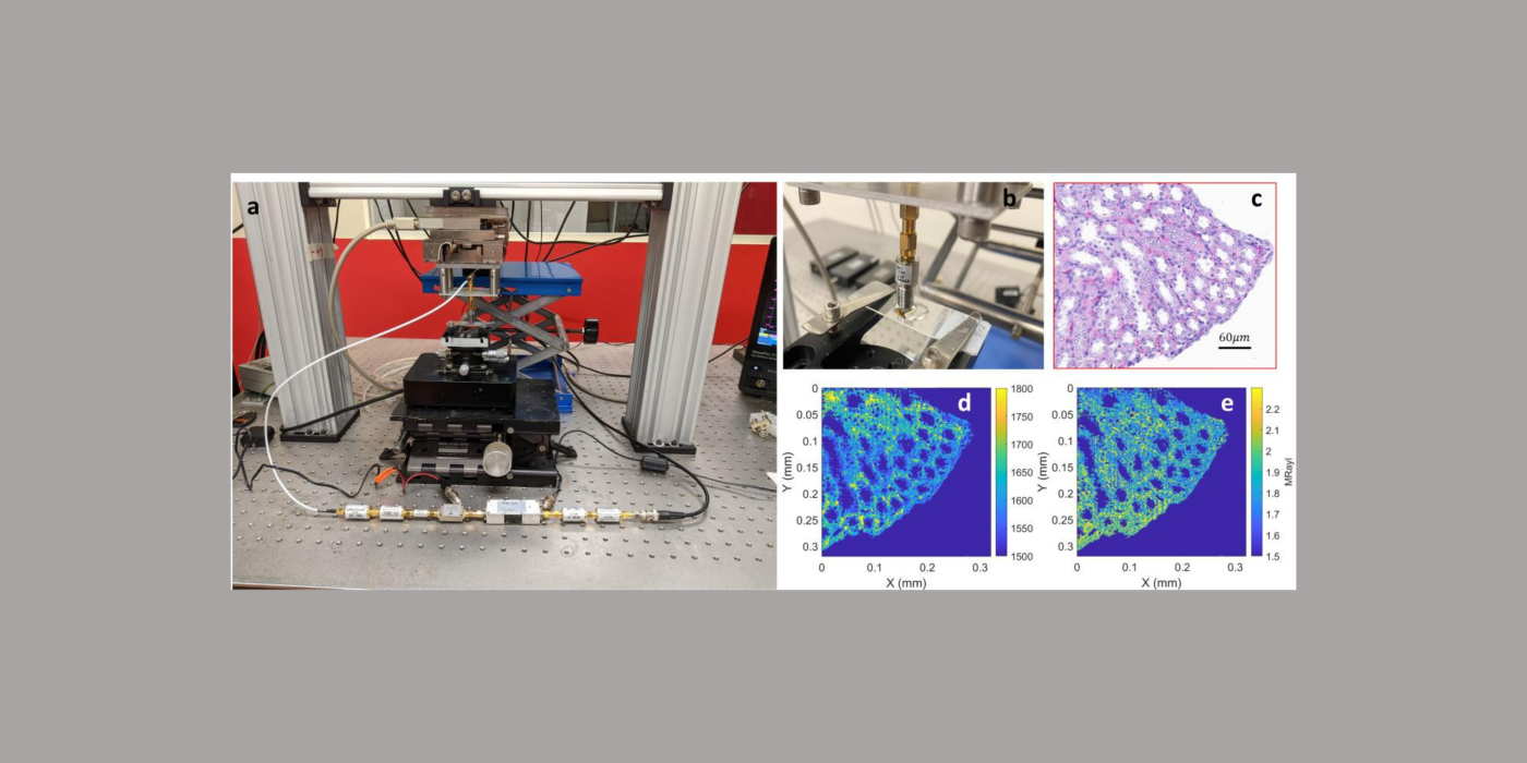
In collaboration with the University of Bristol (Bristol, United Kingdom), and the University of Lyon (Lyon, France), the Mamou lab will develop the next generation of QAM systems. Specifically, data science and coded-excitation approaches will be applied for the first time to QAM technology to yield better image quality, decreased scanning time, and greater ease of use. It will help usher in a new generation of novel, low-cost, user-friendly QAM instruments. QAM permits formation of fine-resolution (i.e., <7 µm at 250 MHz) maps of acoustic and mechanical properties of tissue sections that are <12 µm in thickness. These data can have great value in numerous preclinical investigations. Such property maps are not currently obtainable by any other microscopic-imaging modality, and the new generation of QAM technology—made possible by success in this proposed project—could become widespread in research laboratories and microscopy suites in commercial as well as academic research environments. Such new-generation QAM instruments could be used by technicians with limited knowledge of QAM and, in many ways, their use would be no more complicated than that of a conventional bright-field microscope. Importantly, these novel approaches to QAM will be demonstrated using already available resolution targets, phantoms, and biological tissues (ocular-tissue samples from a guinea-pig model of myopia and cancerous human lymph nodes). During the course of this project, optimal methods will be incorporated in a prototype QAM instrument capable of producing ultra-fine spatial resolution (< 2 µm) images much faster (<1 min) and for a much lower cost than current state-of-the-art QAM systems. In addition, use will be ``turn-key'' (i.e., requiring no technical knowledge and less than one hour of training.)
In-vivo ultrasound-based point-of-care instrument to assess myopia level and progression
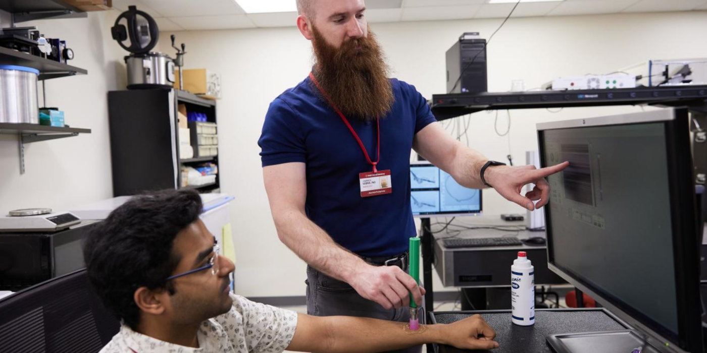
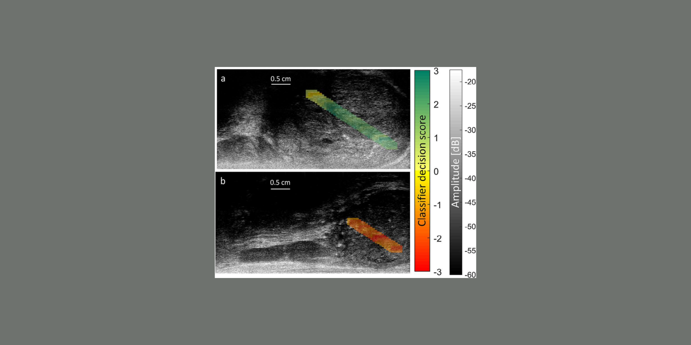
The goal of this project is to develop a novel, low-cost, compact, point-of-care (POC) ultrasound system to assess myopia severity and progression in vivo. Myopia prevalence is increasing worldwide and pathologic myopia is one of the leading causes of blindness. Myopia originates from structural and mechanical changes in the sclera that precede vision-threatening ocular changes. Currently, no method exists to assess myopia progression and severity quantitatively in vivo, and this project seeks to fill this gap. The POC instrument consists of a single-element, spherically focused transducer operating at an 80-MHz center frequency mounted in a handheld probe and connected to a computer equipped with dedicated software, hardware, and a pedal for ease of use implementing a graphical user interface. The POC instrument collects RF, A-line data at a high pulse-repetition frequency while in direct contact with the anterior sclera. The radiofrequency (RF) data is processed using quantitative (QUS) methods to derive so-called QUS parameters associated with the elastic and microstructural properties of the sclera. The QUS parameters are based on the backscatter coefficient and envelope statistics as well as on highly innovative passive elastography. The POC instrument is portable, low-cost, real-time, and nonionizing. It is similar to pachymeters currently used by ophthalmologists for biometric measurements, which will facilitate its adoption by clinicians. Currently, the POC instrument is deployed at our collaborating clinical site, the Singapore Eye Research Institute, to acquire and process data from 100 patients during a prospective clinical study aimed at assessing the value of the POC instrument for evaluating high-myopia patients. Specifically, the clinical study tests the ability of the system to predict myopia severity and progression. If successful, these studies will significantly advance myopia knowledge. Equally importantly, the proposed POC instrument will revolutionize care for myopia patients by providing a low-cost, easy-to-use, and safe means to assess disease severity and progression.
WCM
Outside
Adrian Basarab, Ph.D.
Alin Achim, Ph.D.
Marie Muller. Ph.D.
Stefan Catheline, Ph.D.
Kimberly Showalter Lakin, M.D.
Ronald Silverman, Ph.D.
Donny Hoang, M.D.
Carolyn Bayer, Ph.D.
Jerry Sebag, M.D.
Tadashi Yamaguchi, Ph.D.
Maoxin Wu, M.D., Ph.D.
Kirk Wallace, Ph.D.
The overall goal of this proposed research is to develop and test new ultrasound backscattering models for future quantitative-ultrasound (QUS) -based in-vivo screening and monitoring of early osteoarthritis (EOA). Osteoarthritis (OA) is a joint disease that degenerates articular cartilage (...
The goal of this project is to develop a novel, low-cost, compact, point-of-care (POC) ultrasound system to assess myopia severity and progression in vivo. Myopia prevalence is increasing worldwide and pathologic myopia is one of the leading causes of blindness....
The goal of this project is to develop the next generation of quantitative acoustic microscopy (QAM) systems. Specifically, data-science and coded-excitation approaches are applied for the first time to QAM...
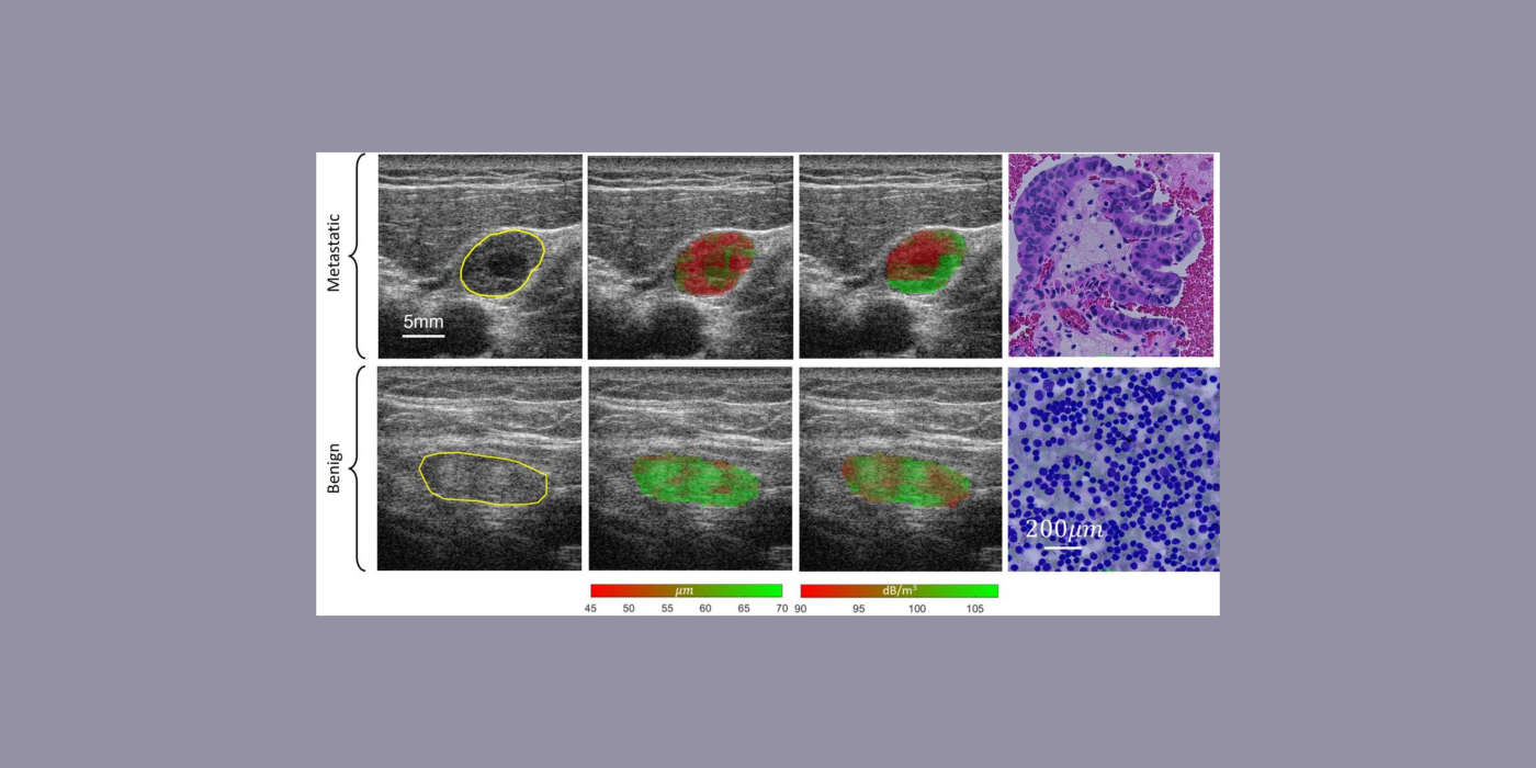
This project addresses the need for reliable, highly sensitive means of detecting metastases to lymph nodes (LNs) and distinguishing them from primary lymphomas and LNs affected by benign conditions. This capability will allow improved staging and treatment of disease. ...