Dr. Kovanlikaya is a neuroradiologist with specific training and expertise in advanced magnetic resonance imaging (MRI) techniques such as MR perfusion, diffusion and diffusion tensor imaging (DTI)—and their applications in brain imaging. Dr. Kovanlikaya has administered several projects with other researchers and published peer-reviewed papers. Dr. Kovanlikaya has collaborated with Dr. Yi Wang over the past 10 years, applying quantitative susceptibility mapping (QSM) imaging techniques to different brain disorders such as brain tumors, multiple sclerosis and intracranial hemorrhages. Recently, his focus has been on Alzheimer’s disease (AD). He is the Co-PI of a funded R-21 grant in which the team proposes to establish QSM as an imaging biomarker for predicting neurodegeneration in AD.
Associated Lab Members

Dr. Kovanlikaya graduated from Hacettepe University, Medical School, Ankara, Turkey (1978), and completed his radiology residency (1982) at the same institution. During his tenure as an attending radiologist in the Department of Radiology at Dokuz Eylul University, Izmir, Turkey, he was appointed Dean of the Medical School in 1999. He also served as professor and chair of the department of radiology, at Yeditepe University, Istanbul, between 2004 to 2007. He joined the department of adiology at Weill Cornell Medicine in 2007.
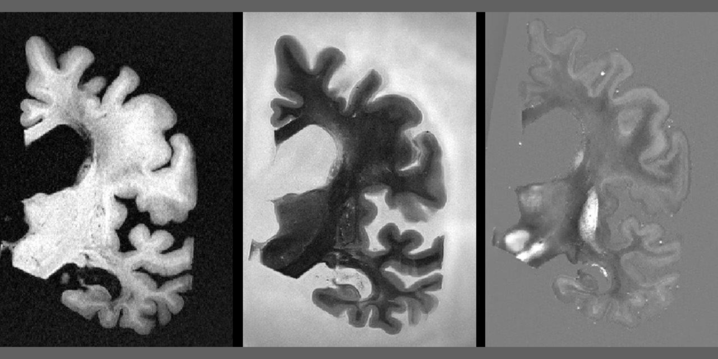
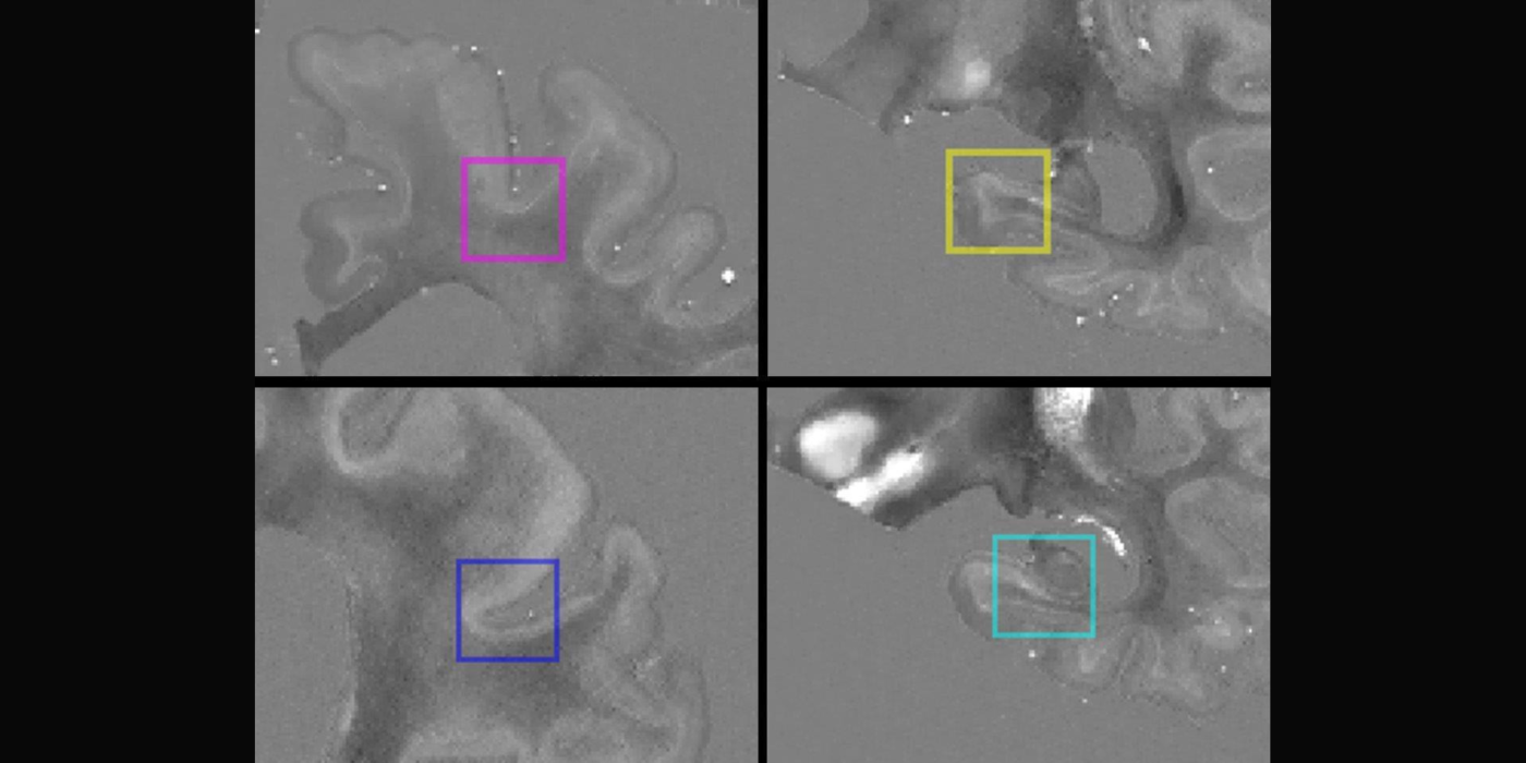
A long-term objective of our research is to establish QSM as a noninvasive MRI marker for predicting neurodegeneration in AD. Our scientific premise is that QSM can measure iron overload involved in AD progression. We propose to...
Award or Grant: The Leon Levy Foundation
In this research project, the Kovanlikaya lab used advanced magnetic resonance imaging (MRI) techniques including diffusion tensor imaging (DTI), arterial spin labeling (ASL), phase c...
Although advanced imaging techniques provide quantitative, metabolic and functional data about brain tumors, it is evident that, as yet, there is no single technique that can offer a complete picture of brain tissue and tumor characterization. By generating a...
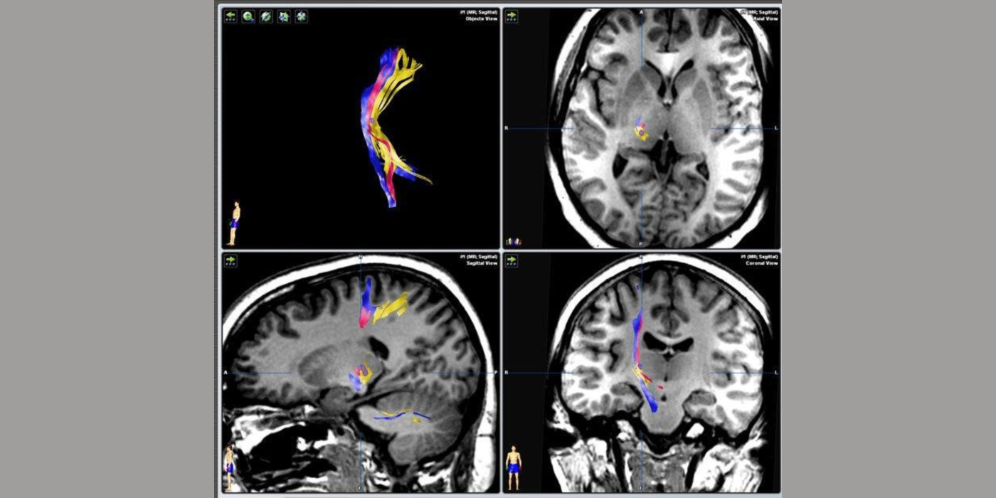
The purpose of this project was to preoperatively identify the thalamic ventroposterolateral nucleus by diffusion tensor imaging (DTI)-fiber tractography and confirm it intraoperatively. A variety of pain syndromes have been treated successfully with deep brain...
The aim of this project was to establish a standard normative database of advanced magnetic resonance (MR) techniques, including but not limited to diffusion tensor imaging (DTI), MR perfusion, and 3D v...
In this research project, the Kovanlikaya group used advanced magnetic resonance imaging (MRI) techniques including diffusion tensor imaging (DTI), arterial spin labeling (ASL), phase contrast cerebrospinal fluid (...