Molecular Imaging Innovations Institute (MI3)

Director: 
Michelle Bradbury, M.D., Ph.D.

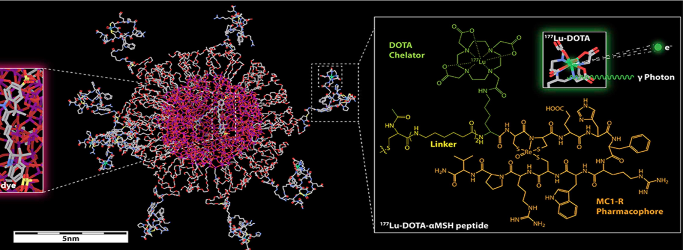
The Molecular Imaging Innovations Institute (MI3) is dedicated to the development of imaging probes and technologies that enable the institute to visualize disease and therapeutics at multi-scale levels. The institute’s collaborative environment serves as a nexus for pre-clinical and clinical translational programs. The MI3’s mission is to drive innovation of new diagnostics and therapies from bench to bedside, and to accelerate the development and translation of personalized medicine. The institute emphasizes translational medicine in oncology, cardiovascular diseases, neurologic and neuropsychiatric sciences, obesity research, and infectious diseases. MI3 is at the forefront of multiple platform technologies including radiochemistry, nanotechnology, polymer and synthetic chemistry, optical imaging, medical physics, and antibody and cell engineering.

Radiotracers

We are developing novel radiotracers to identify diagnostic and prognostic imaging biomarkers for clinical disease management, and to deliver therapeutic radiation for cancer therapy. Examples include positron emission tomography (PET) radioactive tracers to detect early myocardial damage, dysfunctional metabolism, and cancer biomarkers. PET radioisotopes are combined with small molecules, peptides, and biologics such as antibodies for imaging biomarkers.

Drug delivery systems

MI3 investigators utilize multi-scale carriers to deliver drugs specific to diseased cells and tissues. Examples include: 1) small molecules against immune checkpoints to overcome immune suppression, 2) peptide-based nanofibers to achieve shape-controlled tumor uptake, charge-assisted tissue penetration, and enzyme-induced tumor retention, 3) multi-step pre-targeted radioimmunotherapy to improve therapeutic index and pharmacokinetics of therapeutic radioisotopes, and 3) antibody-drug conjugates.

T cell immunotherapy

We are developing T cell immunotherapy against cancers for simultaneous therapy and imaging. Examples include somatostatin receptor-2 (SSTR2) for imaging T cell distribution and activity in vivo using PET/computed tomography (CT), and micromolar affinity chimeric antigen receptor (CAR) T cells to be selective to antigens overexpressed in tumor/tumor stroma while sparing normal cells with basal expression of the same antigen.