![](https://radiology.weill.cornell.edu/sites/default/files/styles/media_album_image/public/wcm_slide_images/razlighi_lab_banner_1.png?itok=zQJKx-gc)
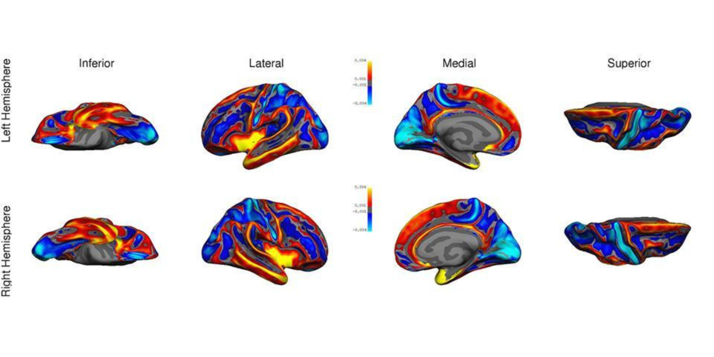
Imaging the hierarchical structure of large scale networks
First principal component of the cortical thickness of 416 healthy participants (20<age<80 years)
![](https://radiology.weill.cornell.edu/sites/default/files/styles/media_album_image/public/wcm_slide_images/razlighi_lab_banner_2.png?itok=4toEWIxy)
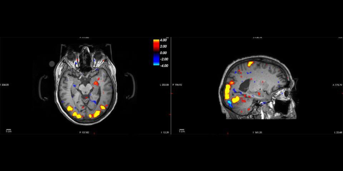
Detecting negative blood oxygen level dependent (BOLD) response in the vicinity of positive BOLD response in the visual cortex due to flashing checkerboard
![](https://radiology.weill.cornell.edu/sites/default/files/styles/media_album_image/public/wcm_slide_images/razlighi_lab_banner_3.png?itok=AkTL_LM-)
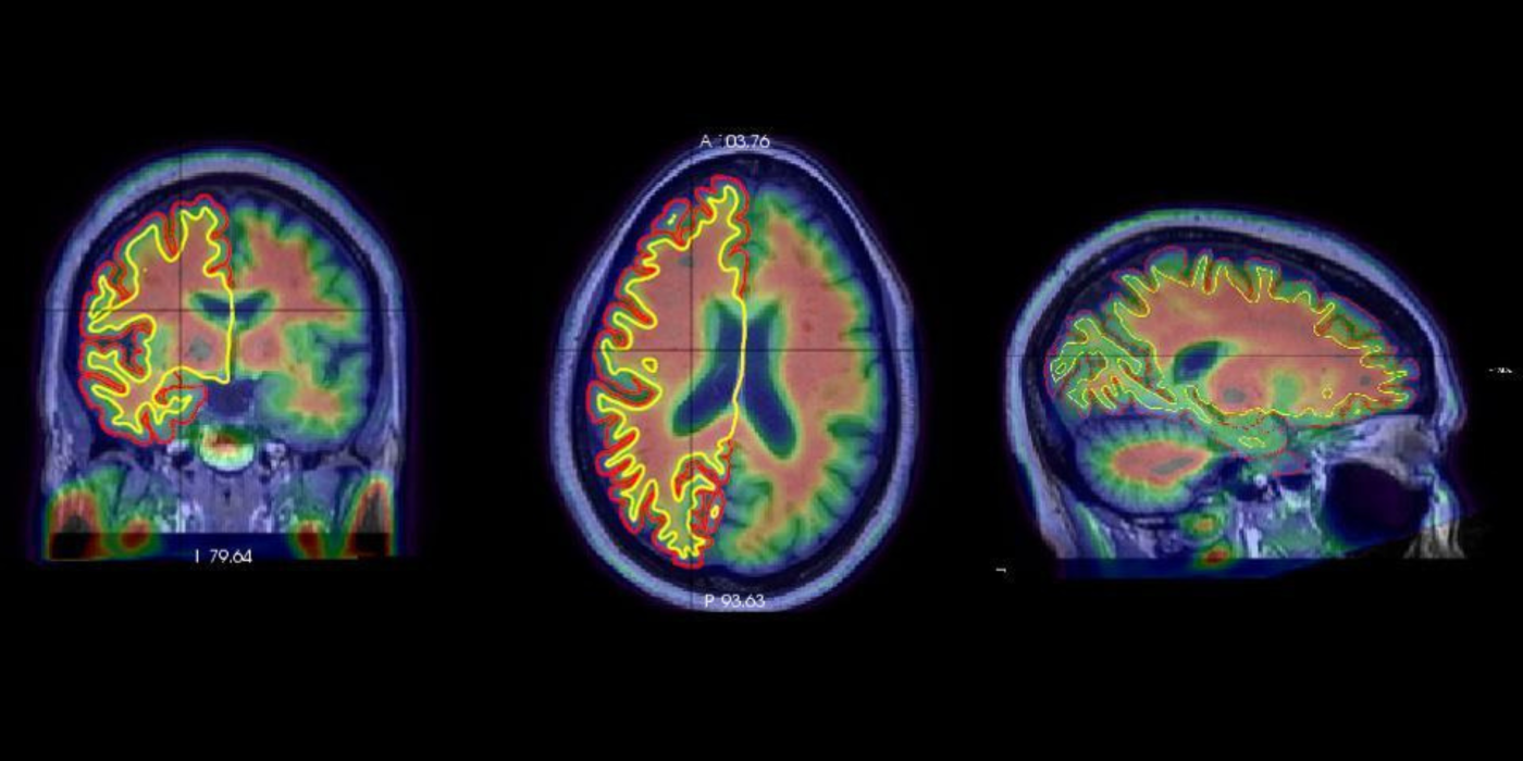
Quantification of amyloid plaques using in vivo positron emission tomography (PET) scanning with florbetaben on the surface of human brain cerebral cortex and white/gray-matter boundary in a healthy participant
![](https://radiology.weill.cornell.edu/sites/default/files/styles/media_album_image/public/wcm_slide_images/razlighi_lab_banner_4.png?itok=-gTrLcyU)
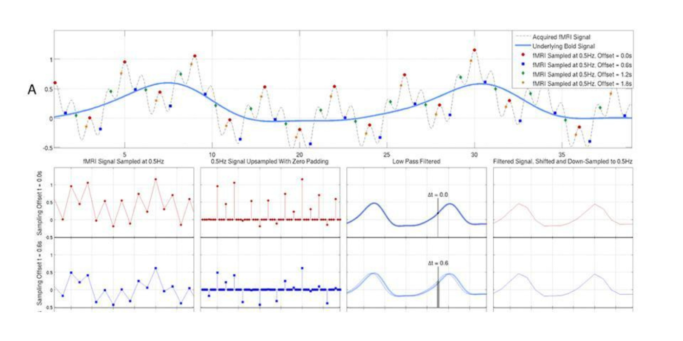
Optimal recovery of blood oxygen level dependent (BOLD) signal
Theoretical illustration of optimal signal reconstruction sampled with different time offset under the Shannon-Nyquist sampling theorem
![](https://radiology.weill.cornell.edu/sites/default/files/styles/media_album_image/public/wcm_slide_images/razlighi_lab_banner_5.png?itok=HKrtD_lB)
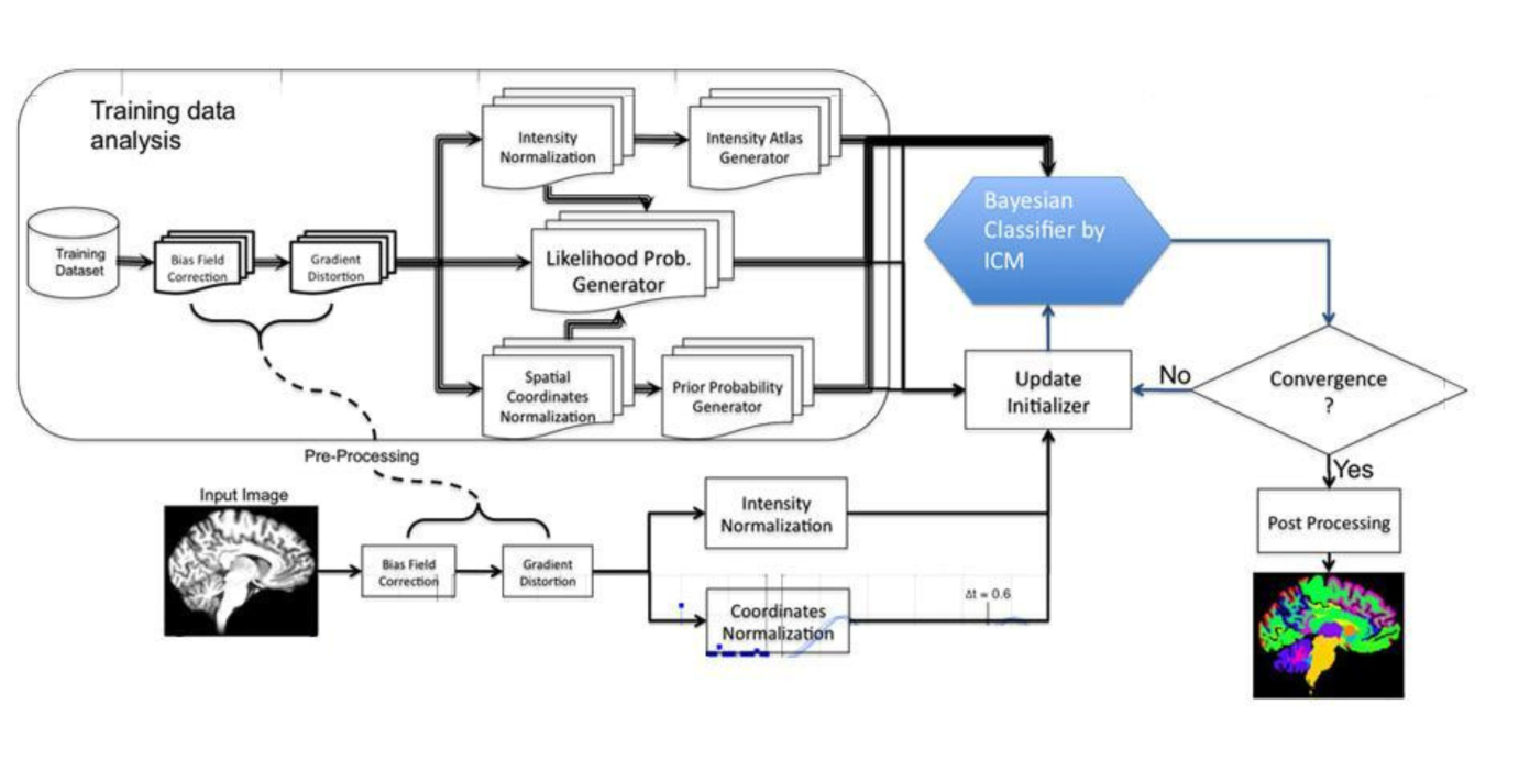
Multilateral Markov random field and brain subcortical segmentation
Flowchart of the pipeline for subcortical segmentation of the human brain using quadrilateral Markov random field and under maximum a posteriori framework
![](https://radiology.weill.cornell.edu/sites/default/files/styles/media_album_image/public/wcm_slide_images/razlighi_lab_banner_6.png?itok=uaxqY7Ul)
![](https://radiology.weill.cornell.edu/sites/default/files/styles/media_album_image/public/wcm_slide_images/razlighi_lab_banner_7_0.png?itok=zMDZbPAF)