Advanced magnetic resonance imaging for the study of normal pressure hydrocephalus
In this research project, the Kovanlikaya group used advanced magnetic resonance imaging (MRI) techniques including diffusion tensor imaging (DTI), arterial spin labeling (ASL), phase contrast cerebrospinal fluid (CSF) flow measurement, and structural volumetric MRI to develop quantitative criteria to diagnose normal pressure hydrocephalus (NPH) patients more precisely.
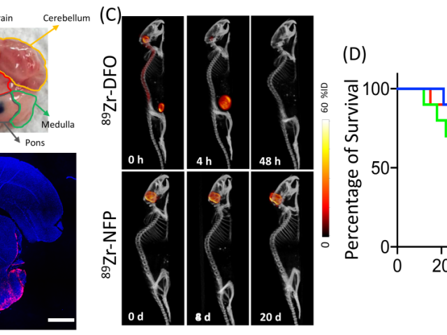
Multifunctional nanofiber for convection-enhanced delivery of theranostics to diffuse intrinsic pontine Glioma (DIPG)
Among all pediatric cancers, DIPG stands out as the most aggressive. Focal radiotherapy only extends patient survival for a few months, and chemotherapy proves ineffective as DIPG inherently resists most chemotherapeutics. Moreover, the blood-brain barrier (BBB) naturally restricts many drugs from reaching the brain tumor. Convection-enhanced delivery (CED), a direct infusion technique for...
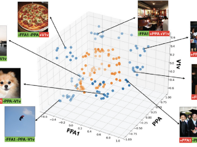
Creating and validating a novel, non-invasive method for targeted brain activation
Neuromodulation (the localized manipulation of neuronal activity) holds therapeutic promise for neurological/neuropsychiatric diseases. There is currently an unmet need for a non-invasive, safe, personalized and inexpensive method of neuromodulation that can be used to functionally target and manipulate brain networks to achieve therapeutic goals. The lab’s long-term goal is to develop a novel...
WCM catchment Prostate Cancer Health Impact Program (pCHIP)
Dean’s Health Disparity Research Award WinnerProstate cancer (PCa) is the second leading cause of cancer death in American men. Several studies have shown higher mortality rates among African American and Hispanic men relative to White men. Lower screening rates in ethnic minorities, along with poor access to treatment resources, medical information, high quality of care, new imaging modalities,...
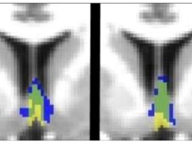
Human septal nuclei
Septal nuclei, located in the anterior basal forebrain, exert strong control over hippocampal function and are critical for memory in animal models, yet remain understudied in humans. Until very recently, septal nuclei were not included in any of the standard neuroanatomic templates commonly used to interpret neuroimaging studies. We therefore developed and validated a new magnetic resonance...
Biomarkers of neurodegeneration in women who have experienced repetitive traumatic brain injury due to intimate partner violence
This pilot project will assess positron emission tomography (PET) and magnetic resonance imaging (MRI) biomarkers of neurodegeneration in female victims of intimate partner violence (IPV) with and without TBI. The goal is to begin to investigate risk Alzheimer's disease (AD) and chronic traumatic encephalopathy (CTE) in this highly vulnerable and medically underserved population. While intense...
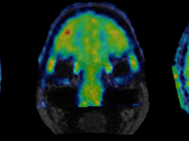
PET imaging of neuroinflammation
Inflammation is a vital, complex process by which certain cells or tissues identified by the immune system as abnormal (e.g., foreign, damaged or dead) can be repaired or removed to facilitate continued survival of remaining cells and the organism. Excessive or dysregulated inflammation can be harmful, especially in the brain We uses Positron emission tomography to assess inflammation in aging...
The Sleep MRI Study
This project monitors brain function changes during sleep, including perivascular space, brain perfusion, cerebral vascular pulsation, and magnetic resonance imaging (MRI)-compatible electroencephalogram (EEG).
CSF Clearance in Sporadic Alzheimer's Disease
Award or Grant: NIH R01 AG057848 (2018-2023)Cerebrospinal fluid (CSF) drainage pathology, a potential new Alzheimer's disease (AD) therapeutic target, has been hypothesized as impaired in AD and demonstrated in AD models. There is no non-invasive technique to measure brain CSF clearance (BCC) in humans. Low molecular weight positron emission tomography (PET) tracers such as 18 fluorodeoxyglucose...
Hypertension, brain clearance, and markers of neurodegeneration
Awards and Awards: R01, National Institutes of Health (NIH)Reduced clearance of brain waste has emerged as a possible factor underlying neurodegeneration. Although it has long been hypothesized that vascular conditions may reduce the brain’s clearance capacity (through reduced cerebral blood flow (CBF), and diminished vascular pulsations), there is no direct evidence in humans supporting the...


