X-Ray & Emission Imaging Laboratory (XEIL)

XEIL Lab Director


“Amir

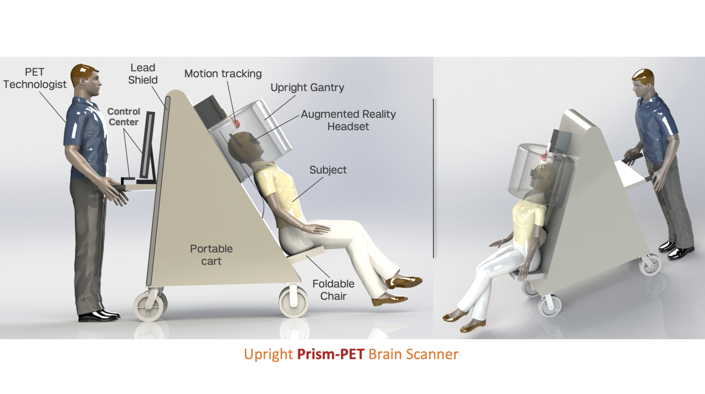
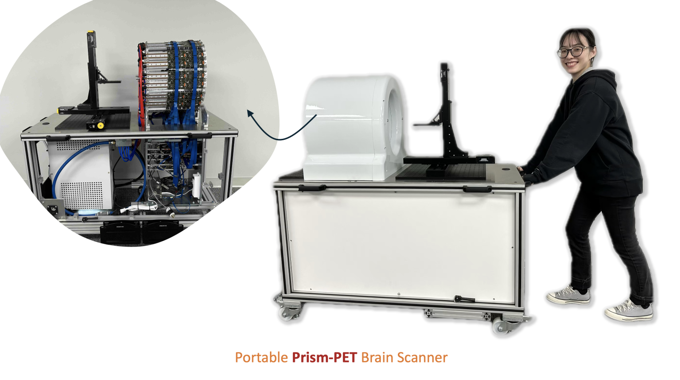
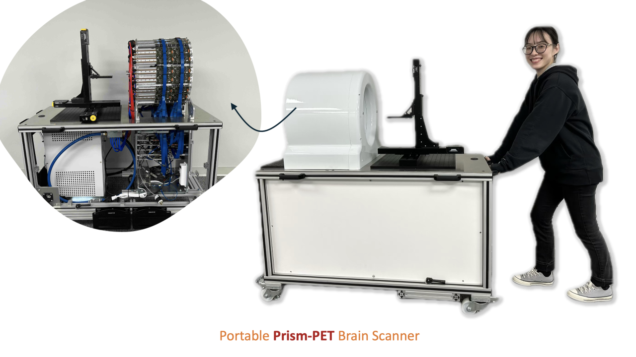
Amir H. Goldan, Ph.D., develops photon-counting x-ray imaging and PET medical imaging detectors and systems. The research of Dr. Goldan, who received his doctorate in electrical and computer engineering from the University of Waterloo, Canada, has been funded by the National Institute of Health (NIH), the National Science Foundation, and the Defense Advanced Research Projects Agency. Recently, the NIH awarded Dr. Goldan a $6.25M U01 grant to develop Prism-PET, a high-performance, compact, portable, upright brain PET scanner featuring motion compensation and CT-less attenuation correction. For clinical translation, Dr. Goldan and the XEIL Lab will use [18F]MK6240 radiotracer, which has subnanomolar affinity for tau neurofibrillary tangles. They will leverage Prism-PET imaging's ultra-high resolution topographical capabilities — such as uptake in small regions like the entorhinal cortex and hippocampus — to perform early-stage Braak staging in asymptomatic individuals with mild cognitive impairment.

XEIL Mission

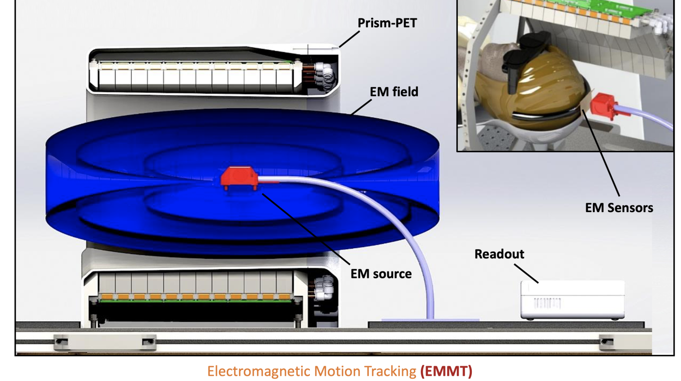
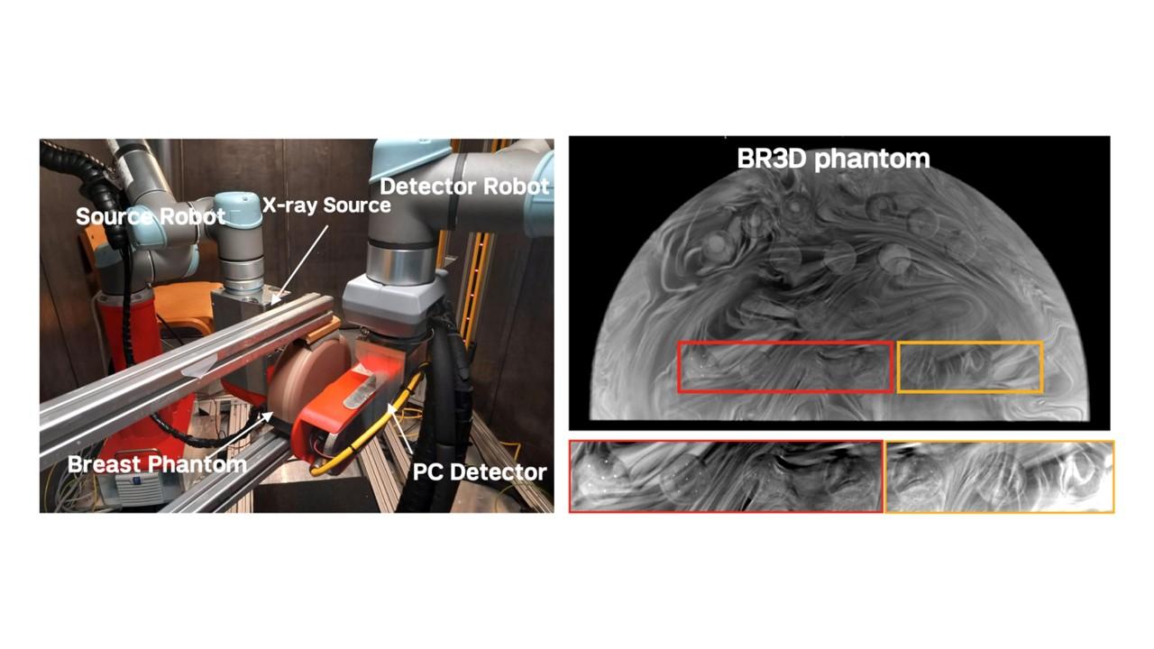
XEIL has over a decade of experience in developing novel multimodal diagnostic systems, quantitative imaging biomarkers, image reconstruction with applications in early-stage Braak staging of Alzheimer’s Disease, behavioral neuroscience, staging of carotid artery plaques, and contrast-enhanced breast imaging. Our work focuses on high-resolution arbitrary path photon-counting computed tomography (HiRAP-PCCT), high-resolution arbitrary path photon-counting mammography (HiRAP-PCM), ultra-high resolution PET, interpretable inverse neural rendering for PET image reconstruction and denoising, motion tracking and correction for PET imaging, functional dynamic PET imaging, immersive augmented reality-based brain stimulation with simultaneous eye tracking, and radiation detector physics and instrumentation. 

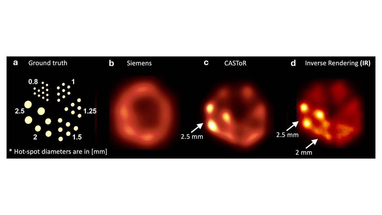
XEIL has developed a prototype Prism-PET brain scanner, achieving the world’s highest resolution PET images of brain phantoms showing reconstruction of 0.8 mm hot spots. Our group also developed a prototype HiRAP photon-counting x-ray imager for single-exposure contrast-enhanced mammography and a novel high-resolution PET image reconstruction platform; the latter utilizes inverse rendering based on physically-based differentiable rendering and gradient-based optimization.