Dr. Ketterling’s lab is focused on the development and translation of ultrasound technology to basic science, small-animal, and clinical applications. He has 20-plus years of National Institutes of Health (NIH)-funded research experience in a variety of ultrasound imaging and therapy topics. Since 2022, he has been at Weill Cornell Medicine (WCM), where he and Jonathan Mamou founded the Biomedical Ultrasound Research Laboratory (BURL).
Dr. Ketterling has worked extensively with high-frequency ultrasound (US) (> 20 MHz). He developed and patented a method for fabricating annular-array transducers. He also designed imaging platforms for ophthalmic human subject use and for studies of in-utero mouse embryonic central nervous system development. The work was extended to in vivo photoacoustic imaging of mouse embryos. The transducer technology was licensed for ophthalmic ultrasound imaging.
More recently, Dr. Ketterling has shifted into a new research area focused on high-speed plane-wave ultrasound imaging. Working with collaborators at New York University (NYU) Medical School and Columbia Irving Medical Center, he applied the technology to several applications including intracardiac flow patterns in mouse embryos and adult mice, blood flow in the front and back of the human eye, and activation of acoustic nanodrops for imaging microcirculation.
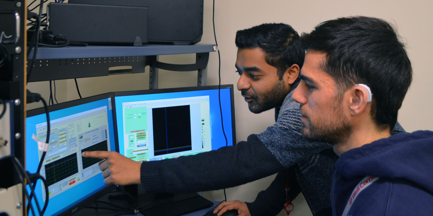
Associated Lab Members

Dr. Jeff Ketterling is a professor of biomedical engineering in radiology at Weill Cornell Medicine. Jeff received his B.S. in electrical engineering from the University of Washington in 1994, and his Ph.D. in mechanical engineering from Yale University in 1999. Jeff’s research focuses on the development and translation of ultrasound technology to basic science, and small-animal and clinical applications, particularly in the realm of high-frequency ultrasound. Recent projects include high-speed plane-wave ultrasound imaging for intracardiac flow patterns in mouse adult mice, blood flow in the front and back of the human eye, and activation of acoustic nanodrops for imaging microcirculation.

Dr. Mamou is a professor of electrical engineering in radiology at Weill Cornell Medicine. Jonathan earned a B.S. in electrical engineering from the Ecole Nationale Superieure des Telecommunications in Paris, France, in 2000. He earned a Ph.D. in electrical engineering in 2005 from the University of Illinois at Urbana-Champaign. His fields of interest include theoretical aspects of ultrasonic scattering, ultrasonic medical imaging, ultrasound contrast agents, and biomedical image processing. Recent projects focus on quantitative ultrasound methods for ultrasound tissue characterization in oncology and ophthalmology, and ultra-high frequencies (>250 MHz) for quantitative acoustic microscopy, among others.

Dr. Cameron Hoerig is a research associate with the Weill Cornell Medicine Department of Radiology Biomedical Ultrasound Research Laboratory (BURL). He received his B.S. from the University of Cincinnati in 2013. He then entered the bioengineering graduate program at the University of Illinois at Urbana-Champaign, where he received his M.S. and Ph.D. degrees in 2015 and 2018, respectively. His research interests include biomedical ultrasonic imaging, image science, computational mechanics, and machine learning. Dr. Hoerig’s current research projects focus on quantitative ultrasound for soft tissue characterization over a broad range of imaging frequencies spanning 10MHz up to 1GHz.

Dr. Geraldi Wahyulaksana, Ph.D., received his MS.c. in electrical engineering from the Eindhoven University of Technology in 2017 and his doctorate in biomedical engineering from Erasmus University Medical Center in 2024. His research focuses on cardiac-blood-flow imaging using high-frame-rate ultrasound, covering both vasculature and chamber studies. Recent projects include plane wave imaging for intracardiac flow patterns in mice and imaging myocardial perfusion with contrast-enhanced ultrasound in porcine.

Ladan Yazdani, Ph.D., received her doctorate in biomedical engineering from the University of Montreal. Before her doctoral studies, she completed both her M.S. in biomedical engineering and B.S. in electrical engineering from the University of Tehran. Her research interests include medical ultrasound imaging, signal and image processing, quantitative ultrasound, and elastography. Her recent projects focus on the development and application of ultrasound techniques, specifically targeting the characterization of homogeneity and inhomogeneity in soft tissues, which holds significant potential in advancing diagnostic methodologies in medical imaging.
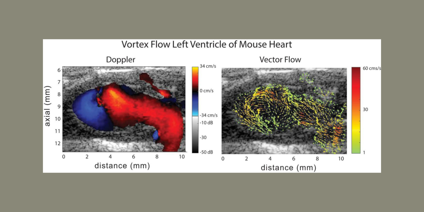
Cardiovascular disease (CVD) accounts for one of every three deaths each year in the U.S. Mice are the most common model organism for translational CVD studies of the mammalian heart. Ultrasound is now extensively used in small animals to obtain cardiac functional parameters. However, advanced ultrasound (US) intracardiac vector-flow imaging techniques that are gaining traction for human CVD, such as cardiomyopathies, have yet to translate to preclinical use, thus limiting the functional cardiac parameters that can be obtained from mice.
This project, in partnership with Dr. Oralkan at North Carolina State University, and Drs. Fishmann and Phoon at New York University (NYU) School of Medicine, seeks to develop a 30-MHz, 2D-capacitive micromachined ultrasonic transducer (CMUT), row-column (RC) high-frequency-ultrasound array, and a plane-wave vector-flow imaging approach capable of sub-ms, full-frame image capture for intracardiac imaging in mice. The CMUT array will let us collect data in adjacent planes to provide a 3D view of flow dynamics within the murine heart. We will study intracardiac left ventricle (LV) blood flow patterns in two highly related mouse strains that display divergent responses (progressive hypertrophy vs. dilatation and failure) to abnormal pressure overload. We seek to quantify abnormal left ventricle flow patterns relative to sham control mice, and to detect flow disruption prior to changes in traditional functional echo or strain measures. The ability to detect subtle phenotypic changes in common mouse models of CVD that are a result of early-stage diseases, or therapies, may translate to earlier and more aggressive treatment of patients at highest risk of pressure-overload induced heart failure.
The goal of this project is to implement quantitative ultrasound (QUS) capabilities into a clinical ophthalmic ultrasound system to characterize vitreous inhomogeneity (i.e., clinically significant vitreous floaters called vision degrading myodesopsia) as it relates to states of health and disease. Age-related changes due to collagen cross-linking and aggregation with liquefaction create inhomogeneities that appear non-uniformly throughout the vitreous body. For patients with myopia, these processes occur earlier in life, when vitreo-retinal adhesion is still strong. They destabilize the vitreous body before adhesion to the retina is weakened, resulting in a variety of conditions that impact vision. The ability to depict vitreo-retinal organization will offer unique early-stage detection, and assessment of vitreo-retinal disease, in patients with myopia at risk for retinal detachment and vitreo-maculopathies resulting from traction.
In collaboration with Quantel Medical, the VMR Institute (Dr. Jerry Sebag), and Columbia University Irving Medical Center (Dr. Ronald Silverman) we seek to incorporate a new 3D probe and QUS capabilities into a state-of-the art, 20-MHz annular-array-based clinical ophthalmic ultrasound system. The initial phase of the project emphasized device enhancement processing of the ultrasound data and collection of age-normal and myopic patient data. The later stages of the project will focus on development and integration of a probe capable of volumetric acquisition, further patient data collection, and QUS classification methods that take advantage of the new volumetric data. The final system will permit quantitative characterization of the vitreous body and allow for data-based treatment decisions of vitreo-retinal diseases.
The goal of this translational project is to add quantitative ultrasound (QUS) capabilities to a clinical ophthalmic ultrasound system to characterize vitreous inhomogeneity (i.e., clinically significant vitreous floaters referred to as Vision Degrading Myodesopsia) as it relates to...
Cardiovascular disease (CVD) accounts for one of every three deaths each year in the U.S. A substantial proportion of patients with CVD develop myocardial dysfunction. Imaging tools that permit early detection of abnormal...
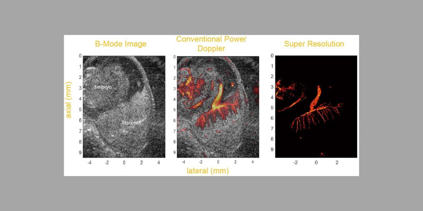
A fundamental question in developmental biology is how specific molecules and genetic pathways control morphogenesis during embryonic development. Recent studies have shown that many of the same molecules and genetic pathways affecting organogenesis are involved in vascular development and patterning during...
Glaucoma is one of the primary causes of irreversible vision loss in this country and worldwide. It is a multi-factorial disease, or family of diseases, characterized by death of retinal ganglion cells (RGCs) and optic...
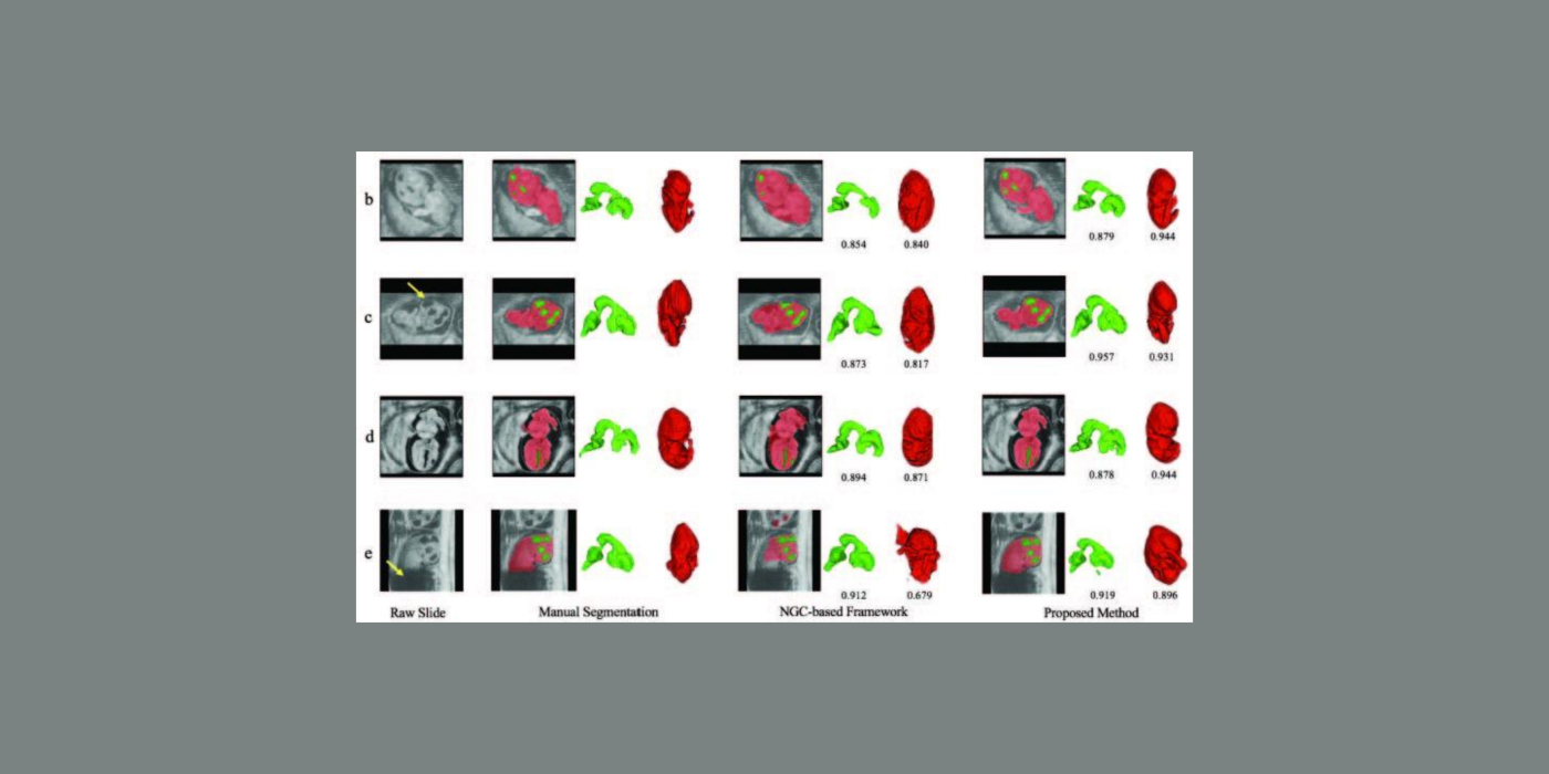
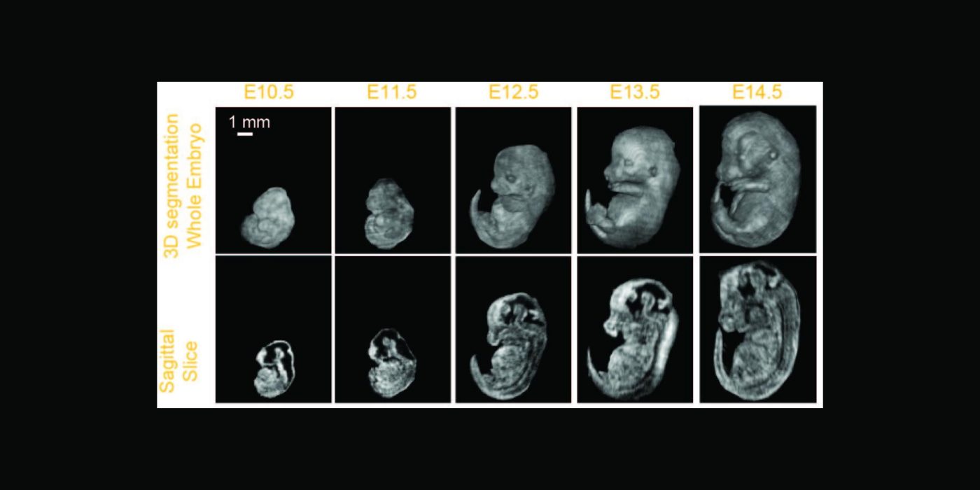
The goal of this proposal: phenotype early- to mid-gestational mouse embryos by segmenting select organ systems in 3D data sets acquired in utero with high-frequency...
Glaucoma is an optic neuropathy and the leading cause of blindness, affecting over 3 million people in the United States. Elevated intraocular pressure (IOP) is a well-known risk factor for this disease, but reduced ocular perfusion is also now appreciated to be an important risk...