
MRSaiFE: Real-time prediction of tissue heating in MRI
A crucial safety concern for ultra-high field (UHF) magnetic resonance imaging (MRI) is the significant radiofrequency (RF) power deposition in the body in the form of local specific absorption rate (SAR) hotspots, leading to dangerous tissue heating/damage. This work is a proof-of-concept demonstration of artificial intelligence (AI) based real-time MRI safety prediction software (MRSaiFE)...

Diffusion MRI of treatment response for de-escalation of radiation therapy
Awards or Grants: UG3/UH3CA228699, National Cancer Institute NCI, 5/1/2019 to 4/30/2024Description: Quantitative diffusion magnetic resonance imaging (dMRI) remains challenging as dMRI data represent different biophysical properties of tissue depending on diffusion weighting strength (q) and diffusion time (t) used for the measurement. The scientific premise of the study is that it will establish...
")
Magnetic resonance guided focused ultrasound (MRgFUS)
Magnetic resonance (MR)-guided focused ultrasound (MRgFUS) is a non-invasive therapeutic modality for neurodegenerative diseases that allows real-time imaging of targeted regions. However, MR image quality is poor and severely limits the technology due to the use of the body coil for focal targeting. Acoustic simulations demonstrated the acoustic transparency (signal loss <1%) of an ultra-...
Stretchable receive array based on liquid metal technology
Magnetic resonance imaging (MRI) relies on a dense array of radiofrequency (RF) coils to obtain functional and anatomical information inside the body. Tightly fitting coil arrays boost the signal to noise ratio (SNR) and imaging speed. Unfortunately, most commercial RF coils are rigid, and of fixed size, as they are intentionally designed to fit the general patient population and hence provide...

A 2D high-pass ladder RF coil architecture for UHF MRI
The Winkler Lab has a dedicated radiofrequency (RF) team with a top-notch RF lab to design, demonstrate and test high field and UHF receive and transmit coils. Our team is devoted to cutting-edge research studies that explore the benefits of ultra-high field magnetic resonance imaging (UHF MRI), and that contribute to clinical imaging practices. Specifically, the lab’s studies focus on novel coil...

Human connectome mapping using ultra-high-resolution MRI: A technological pathway
A great challenge of modern biomedical science is the mapping of the human brain to understand underlying functionality and behavior. The National Institutes of Health (NIH)-funded Human Connectome Project (HCP) is a large-scale, multi-institutional effort building a vast in-vivo database of neural connectivity pathways, acquired using diffusion and functional magnetic resonance imaging (MRI)....
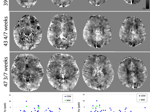
Assessment of brain development in infants using QSM
The early postnatal period is a highly important phase of rapid and dynamic brain development. The lab is investigating regional magnetic susceptibilities in the brains of infants in the first two months of life to relate these measures to developing brain myelination and iron deposition. The lab is also measuring magnetic susceptibilities in infants diagnosed with congenital heart disease in...

Development of non-invasive placental perfusion imaging using velocity-selective ASL (VSASL)
The placenta is a vital organ for transferring oxygen and nutrients from the mother to fetus during pregnancy. Early in pregnancy, the feeding arteries dilate five to 10 times to support rapid fetal growth and development. When this remodeling is incomplete or fails, blood flow is not adequately supplied to the fetus: placental insufficiency occurs. So placental perfusion imaging may provide an...

Investigation of regional cerebral blood flow (CBF) in very preterm infants using ASL
While the third trimester of gestation is a crucial phase of rapid brain development, little has been reported on the trajectories of pre-term infant cerebral blood flow (CBF) during this period. When arterial spin labeling (ASL) is applied to newborns, several pitfalls must be avoided when selecting parameters for data acquisition and data analysis. In this study, the lab addressed obstacles to...
 images (A,D), kw maps (B,E), and cerebral blood flow (CBF) maps (C,F) obtained from a 32-year-old subject with no vascular risk factors (A-C), and a 77-year-old subject with one vascular risk factor (former smoker, 40 pack-year) (D-F)")
Quantitative permeability mapping of water exchange across the blood-brain barrier in normal aging and AD
Award or Grant: R01 AG077594-01 (not yet funded)The team’s objective is to develop quantitative permeability mapping (QPM) of kW, the water exchange rate across the blood-brain barrier (BBB), for the detection and quantification of BBB impairment in normal aging and Alzheimer’s disease (AD). Dynamic contrast-enhanced (DCE) magnetic resonance imaging (MRI) can be used to map BBB permeability to an...