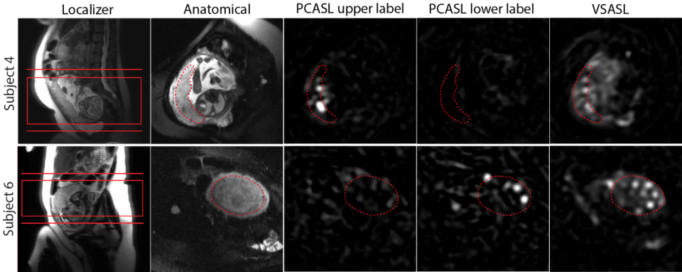
The placenta is a vital organ for transferring oxygen and nutrients from the mother to fetus during pregnancy. Early in pregnancy, the feeding arteries dilate five to 10 times to support rapid fetal growth and development. When this remodeling is incomplete or fails, blood flow is not adequately supplied to the fetus: placental insufficiency occurs. So placental perfusion imaging may provide an early biomarker of placental dysfunction. In this study, the lab developed placental perfusion imaging in vivo using velocity-selective arterial spin labeling (VSASL) and 3D image readout. The lab demonstrated the feasibility of placental perfusion imaging in healthy pregnancies, and showed differences in both global and regional placental perfusion between pregnancies complicated by fetal heart disease and healthy pregnancies.


