The Quantitative Neuroimaging Laboratory (QNL), an engineering-based research lab housed in the Brain Health Imaging Institute (BHII) of the Weill Cornell Medicine (WCM) Department of Radiology, primarily investigates the neural and neurophysiological mechanisms underpinning the negative blood-oxygenation-level dependent (BOLD) response (NBR) in functional magnetic resonance imaging (fMRI) signals. Additionally, the QNL employs NBR as a new tool to study the structure and dynamics of the brain’s large-scale networks and subsystems. By using state-of-the-art signal and image-processing tools, and mathematical and statistical methods, the QNL develops techniques to quantify structural and functional brain images—thereby detecting brain-based effects that are normally beyond the sensitivity and specificity of common detection methods.
Associated Lab Members

In 2009, Dr. Qolamreza "Ray" Razlighi earned his doctorate in electrical engineering and image processing from the University of Texas. During this time, Dr. Razlighi introduced a new causal Markov random field (MRF) model—Quadrilateral MRF (QMRF)—which has dramatically influenced medical and commercial image analysis, resulting in 12 publications and one patent to date. Dr. Razlighi’s advanced knowledge of neuroimaging and neuroscience started with one year of postdoctoral training at the Brain Imaging Laboratory of the Molecular Imaging and Neuropathology Division, New York State Psychiatric Institute, Columbia University. His education continued with two years of postdoctoral training in the Cognitive Neuroscience Division, Department of Neurology, Columbia University.
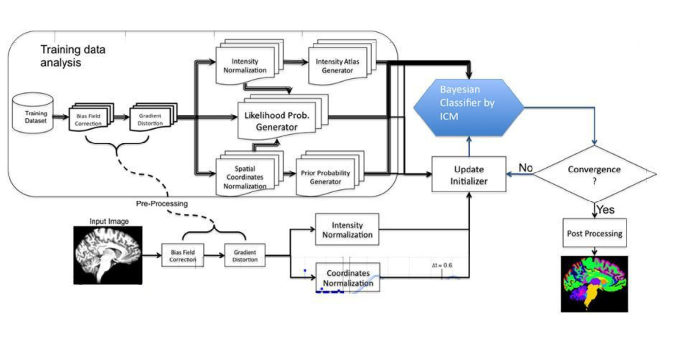
Dr. Razlighi has been involved in many neuroimaging projects focused on implementing mathematical models and methods for magnetic resonance imaging (MRI) and functional magnetic resonance imaging (fMRI) data analysis. These include the development of a method for extracting brain features related to the cortical folding pattern; the development of a new non-stationary maximum a posteriori (MAP) QMRF (MAP-QMRF) classifier for brain image segmentation; analysis of fMRI data in a subject’s native space (thereby circumventing the problematic spatial-smoothing step, particularly in studies comparing young/old); and the investigation of inter-hemispheric averaging in resting-state fMRI data analysis. During his postdoctoral training, Dr. Razlighi participated in numerous neuroscience and neuroimaging courses.

Hani Hojjati, Ph.D., is a postdoctoral associate in the Weill Cornell Medicine Department of Radiology. In 2011, Dr. Hojjati earned his bachelor’s degree in electrical engineering from the Mazandaran University of Iran; from 2011 to 2013, he earned his master's degree in electrical engineering from the Babol Noshirvani University of Technology (NIT); and from 2014 to 2018, he continued his education at NIT, earning a doctorate in electrical engineering.
At NIT, Dr. Hojjati’s thesis focused on predicting Alzheimer's disease (AD) using multimodal neuroimaging methods and machine learning approaches. By employing neuroimaging and multimodal forecasting techniques, he determined accurate predictors for identifying mild cognitive impairment (MCI) versus AD. Using resting-state functional and structural magnetic resonance imaging (MRI) as tools, he enhanced the accuracy of predicting AD.
In 2019, Dr. Hojjati started one year of postdoctoral neuroimaging training at the University of Tennessee Health Science Center Department of Pediatrics. In 2020, he entered a second postdoctoral training program at the Weill Cornell Medicine Department of Radiology. His current research involves using multimodal neuroimaging methods, including structural MRI, and amyloid/tau positron emission tomography (PET), to understand the association between neurodegeneration and amyloid/tau pathologies in the brains of both healthy control and MCI subjects. An award-winning researcher, Dr. Hojjati has published more than 12 journal articles and book chapters and presented at 19 conferences.

A graduate of Daemen College, Sindy Ozoria modeled her individualized studies degree after the history and philosophy of science program at the University of California, Los Angeles. Ozoria, an assistant research coordinator, is interested in applying translational science to vulnerable populations using computational, cognitive, and theoretical neuroscience tools.
By using the NBR as a new tool, the QNL disentangles spatiotemporal discrepancies between well-documented Alzheimer’s disease (AD) pathologies, thereby proposing refinement to the existing hypothesis for AD’s underlying pathophysiology. To achieve these goals, the QNL develops techniques to quantify structural and functional brain images, detecting brain-based effects that are normally beyond the sensitivity and specificity of the field’s commonly used methods.
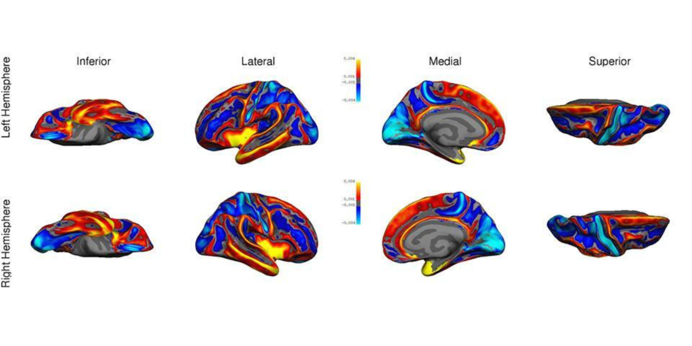
The default mode network’s (DMN’s) topography can be obtained using two different functional magnetic resonance imaging (fMRI) techniques: 1) spontaneous, but organized, synchrony in the low-...
Award or Grant: National Institutes of Health/National Institute on Aging (NIH/NIA) 5R01AG055299, NIH/NIA 5R01AG055422, NIH/NIA 5 R01 AG057681
Conventional Markov random field (MRF) is hampered by...
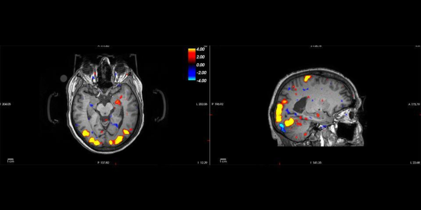
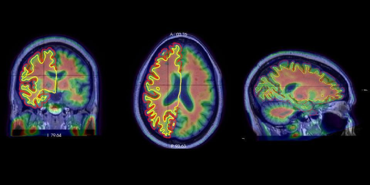
In functional neuroimaging studies of cognitive aging, age-related changes in brain morphology make it difficult to co-register brains, which is a key step for studies comparing task-related activation in young and old groups. To demonstrate the problem’s severity, the video below shows 29 participants’ brains...
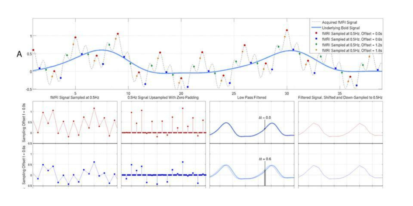
The QNL has developed an optimal technique to extract the blood-oxygen-level-dependent (BOLD) signal from interleaved functional magnetic resonance imaging (fMRI) data. In this project, the lab uses a well-established,...
Award or Grant: National Institutes of Health (NIH)/National Institute on Aging (NIA) 5R01AG026158
Functional magnetic resonance imaging (fMRI) pre-processing requires urgent attention, particularly given the increasingly complex derivation of functional ...
Award or Grant: National Institutes of Health (NIH)/National Institute on Aging (NIA) 3RF1AG038465
A comprehensive functional magnetic resonance imaging (fMRI) simulator not only helps evaluate and improve the present blood oxygenation-level-dependent (BOLD) extraction methods for false-...
The Quantitative Neuroimaging Laboratory’s (QNL’s) main research project is investigating the neural and neurophysiological mechanisms underpinning the negative blood-...