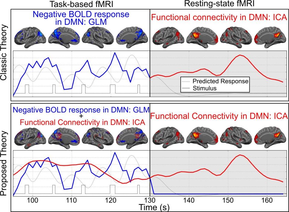
The default mode network’s (DMN’s) topography can be obtained using two different functional magnetic resonance imaging (fMRI) techniques: 1) spontaneous, but organized, synchrony in the low-frequency fluctuations of resting-state fMRI (rs-fMRI), known as “functional connectivity”; 2) consistent, robust deactivations in task-based fMRI (tb-fMRI), here referred to as the negative blood-oxygenation-level dependent BOLD response (NBR).
Based on topographic similarity, these two methods are fundamentally different; however, their results have been used interchangeably to reflect the brain’s resting state, baseline, or intrinsic activities. In the field, the current consensus is that they both represent the same neurophysiological processes; however, recent evidence suggests that these two fMRI techniques measure separate, but overlapping, processes (see figure). For example, the lab's preliminary results suggest that the spatial and temporal expression of the DMN’s functional connectivity remains intact during task performance and is equivalent to the expression of the functional connectivity at rest. Other groups report comparable findings. Also, the lab imposed an alteration in the task-based NBR in the DMN regions by shifting cognitive attention from one sensory stimulus to another; however, this manipulation caused no changes to the DMN’s underlying functional connectivity.
In the DMN regions, both functional connectivity and the NBR have been reported to be disrupted with normal aging, and as a part of the pre-clinical stage of Alzheimer’s disease (AD), mild cognitive impairment, and AD. But in the field, the assumption is that the DMN’s functional connectivity and the NBR represent the same underlying neurophysiological process; meaning, there has been no study, to the lab's knowledge, investigating the cascading events in which functional connectivity or NBR get disrupted.
The lab hypothesizes that a hierarchy of functional subsystems executes brain activities, causing functional connectivity to represent a lower-level process than the task-based network of the NBR.
Functional connectivity networks represent the processes that provide the underlying infrastructure for the functional subsystems represented by the task-based network of the BOLD response. If this is the case, a disruption in the task-based BOLD response shouldn’t necessarily be accompanied by an alteration in the underlying functional connectivity (as the lab has shown in preliminary data); however, any disruption in the functional connectivity must result in an alteration in the task-based BOLD response. The lab aims to test its hypothesis using three experimental designs.
Related Publication
Coming soon.
Software Released
A Python code will be shared upon completion.