Award or Grant: Radiological Society of North America (RSNA) Research and Education Foundation Roentgen Resident/Fellow Research Award
Residents and fellows rotating through the Ballon lab have developed methods for imaging specimens using magnetic resonance imaging (MRI) at high spatial resolution. In one project, the lab achieved high-contrast, high-resolution MR microscopy (MRM) of murine tissue using a 3.0 tesla (T) imaging system. The lab employed a threefold strategy that included customized specimen preparation to maximize image contrast, 3D data-acquisition to minimize scan time, and custom radiofrequency resonator design to maximize signal sensitivity. The lab quantitatively validated the methods through comparisons of neuroanatomy across two lines of genetically engineered mice. The overall methodology is straightforward to implement on clinical MRI systems, and provides ready access to basic MRM at field strengths widely available in both laboratory and clinic.
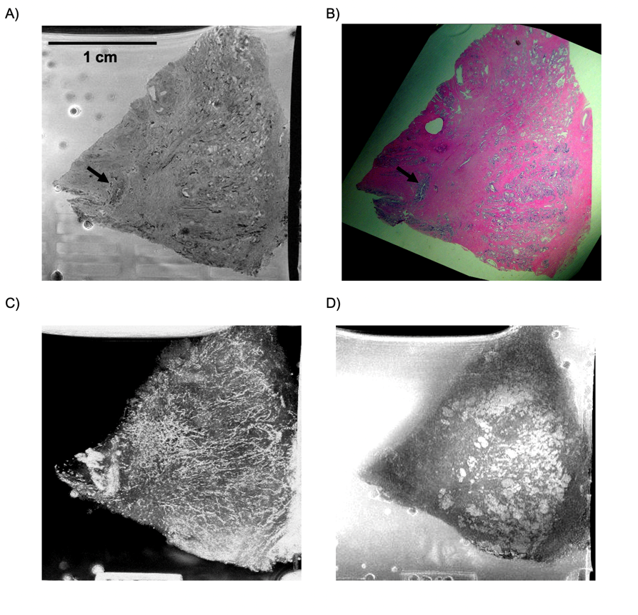
In a second project, the lab determined MR imaging protocol parameters for characterization of prostate tissue at histological length scales. The lab showed that critical histopathological features of the prostate gland can be identified with high-resolution ex vivo MRI examination, and this offers promise that MRI microscopy of prostate cancer (PCa) will ultimately be possible in vivo. As MRI does not require the thin sectioning necessary for optical imaging, the lab could generate both MR micro-angiograms and MR ductograms of prostate tissue for the first time. This work was performed at both 3.0 T and 7.0 T field strengths.
In a third project, the lab undertook a study of breast tissue via MR microscopy. Traditional pathologic evaluation of breast specimens requires a fixation and staining procedure of at least 12 hours, delaying diagnosis and postoperative planning. The Ballon lab introduced an MRI technique using its custom-designed radiofrequency resonators for imaging breast and lymph tissue with sufficient spatial resolution and speed to guide pathologic interpretation and offer value in clinical decision making. In this study, the lab demonstrated the ability to image breast and lymphatic tissue at 7.0 T achieving a spatial resolution of 60 x 60 x 95 µm3 with a signal-to-noise ratio of 15-20, in an imaging time of about one hour. These are the first MR images to reveal characteristic pathologic features of both benign and malignant breast and lymph tissue, some of which were discernible by blinded pathologists who had no prior training in high-resolution MRI interpretation.
Full Caption: (A) 7.0 T gradient echo image of a specimen obtained from organ donor. Image obtained from a 3D acquisition at an isotropic spatial resolution of 60 X 60 X 60 mm3 in 8.5 hours. At this resolution, it is possible to discern specific aggregations of cells (arrows) as confirmed in, (B) the corresponding hematoxylin and eosin (H&E) stained slide. Dark striations are suggestive of microvasculature, which, when processed as a minimum intensity projection image through an entire stack of 40 slices obtained from the 3D gradient echo, produced the angiogram shown in (C). D) Conversely, a maximum intensity projection through the same stack provides a ductogram.


