Amy Kuceyeski Laboratory

Computational Connectomics (CoCo) Laboratory

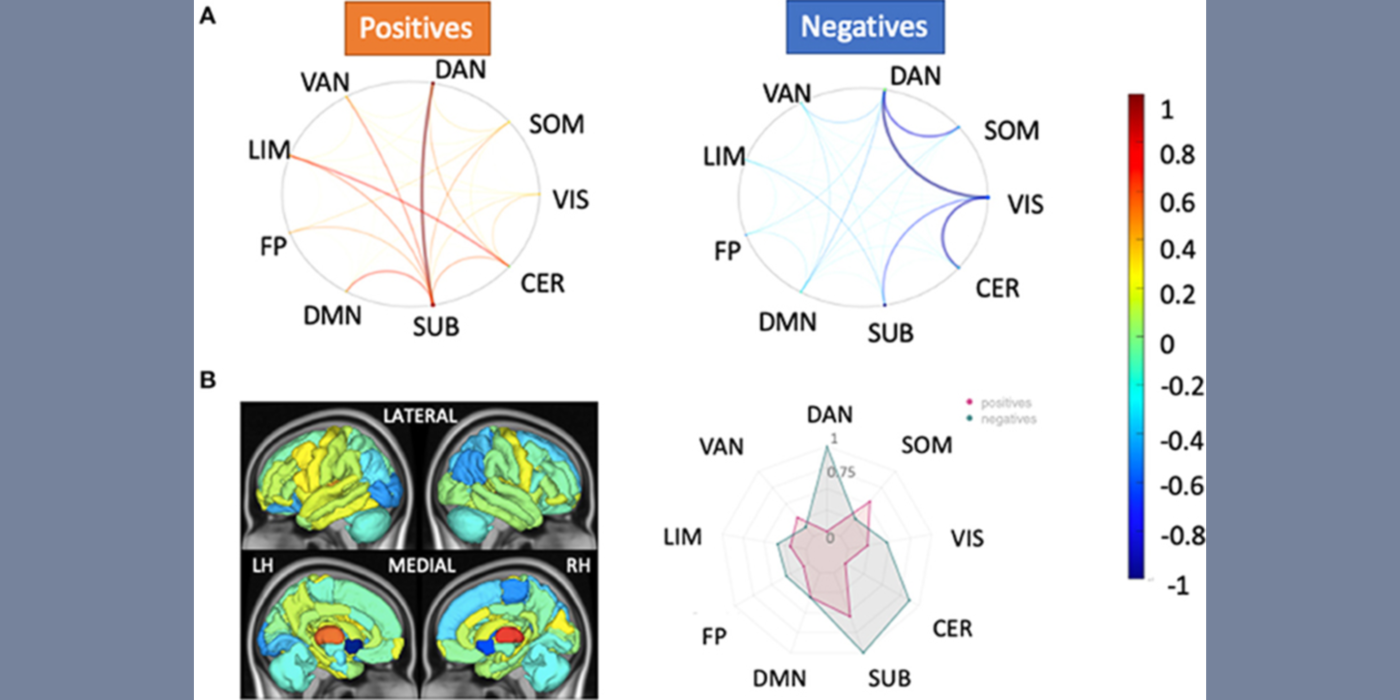
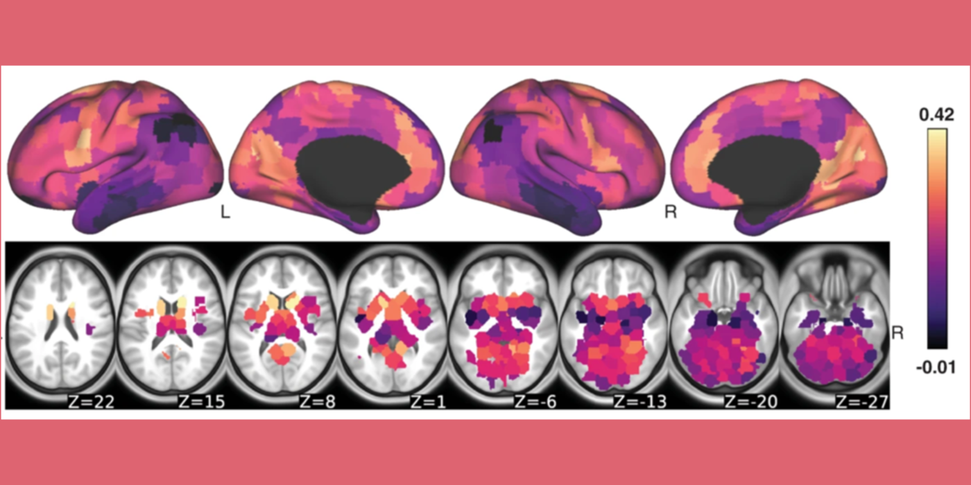
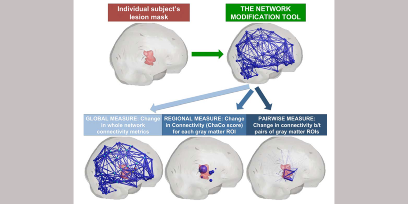
Dr. Amy Kuceyeski’s Computational Connectomics (CoCo) Laboratory is primarily part of the Weill Cornell Medicine (WCM) Department of Radiology, but has secondary affiliations with the Department of Computational Biology on Cornell University's main campus. Her lab focuses on using quantitative methods, including machine learning, applied to neuroimaging data (mostly magnetic resonance imaging (MRI)) to understand the mysteries of the human brain. One major interest of her lab is in uncovering mechanisms of impairment and recovery after neurological injury or disease, including traumatic brain injury, multiple sclerosis (MS) and stroke. Better understanding of brain-behavior relationships will lead to the development of more accurate diagnostics, prognostics and individualized therapeutics that can boost recovery after neurological disease or injury.

Associated Lab Members

View Bio
Amy KuceyeskiPh.D.
  • Professor of Mathematics in Radiology

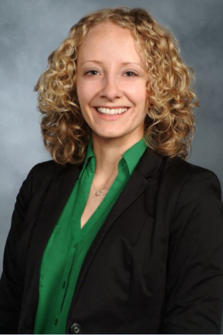
For more than a decade, Amy Kuceyeski has been interested in understanding how the human brain works to better diagnose, prognose and treat neurological disease and injury. Quantitative approaches, including machine learning, applied to data from rapidly evolving neuroimaging techniques, have the potential to enable groundbreaking discoveries about how the brain works. Amy is particularly interested in non-invasive brain stimulation and pharmacological interventions, like psychedelics, that may modulate brain activity and promote recovery from disease or injury. Amy is also the founder and co-director of the cross-campus working group Machine Learning in Medicine, which aims to bring together researchers in Cornell-Ithaca/Cornell-Tech and clinicians/researchers at Weill Cornell Medicine to address medicine's toughest problems. See the group's website. 

Ke HuangPh.D.
  • Research Associate in Radiology
View Bio
Keith JamisonM.S.
  • Staff Associate in Radiology

Keith Jamison is a staff associate in the CoCo Lab. He has a B.S. in computer science and an M.S. in Biomedical Engineering from Cornell University. Through his education and training, he has developed the broad range of skills and expertise necessary to discern scientifically and clinically relevant patterns from large neuroimaging datasets. While working for the Human Connectome Project (HCP) at the University of Minnesota, he helped implement and adapt preprocessing and analysis pipelines for a large number of anatomical, functional and diffusion magnetic resonance imaging (MRI) scans. He also helped design and test new scanning protocols and modalities for some of the HCP-related studies whose data they now propose to analyze. Since joining the CoCo lab in 2017, he has built upon this expertise in neuroimaging acquisition and processing to help develop modeling approaches using this neuroimaging data to better understand the functional and structural connectivity relationship, and how connectivity relates to both healthy brain function and neurological damage or disease. 

View Bio
Sneha PandyaM.S.
  • Research Associate in Radiology

Sneha Pandya is a research associate in biomedical engineering. Over the past eight years, Sneha has worked closely with RajLab, Weill Cornell Medicine (WCM) Multiple Sclerosis Center, and Sumit Niogi’s WCM Lab, serving both radiology and neurology departments, while applying problem-solving techniques to current clinical problems in the imaging, diagnosis and treatment of major brain diseases. The CoCo Lab’s initiative to use quantitative methods and machine learning on multi-modal neuroimaging data to map brain-behavior relationships has inspired her to be part of this lab. The predominant drive of Sneha’s academic career has been to apply these techniques to current issues in neuroimaging. Sneha plans to expand her research pursuits by developing quantitative and machine learning models in understanding structural-functional relationships and predicting early onset of varying brain diseases. 

View Bio
Ceren TozluPh.D.
  • Instructor of Mathematics in Radiology

Ceren Tozlu is a post-doctoral associate in the Weill Cornell Medicine (WCM) Department of Radiology, and in the Cornell University Department of Statistics and Data Science and Computational Biology. She received her M.S. and Ph.D. from the Biostatistics, Biomathematics, Bioinformatics and Health department (3B-H) of Université Claude Bernard Lyon 1 in 2014 and 2018, respectively. She gave lectures to the M.S. students of Cancer, Neuroscience, Biostatistics and Public Health at Université Claude Bernard Lyon for one year and to the medical students at École Santé des Armées (Army Health School of France) for four years. Her M.S. research project focused on application of various machine learning methods to voxel-based conventional human imaging data to predict infarction risk of 3D brain tissue in acute stroke patients. Her Ph.D. thesis focused on modeling disease evolution of stroke and multiple sclerosis (MS) patients based on cross-sectional and longitudinal clinical and imaging data plotted over five years. Her post-doctoral research focuses on (1) modeling of evolution in neurological diseases, particularly in patients with stroke and MS, using statistical and machine learning methods based on demographic, clinical, regional and pair-wise functional and structural connectivity measurements, and (2) identification of the best biomarkers of disease evolution including the particular structural and functional connections contributing to differences in patients with different neurological diseases. Her post-doctoral study aims to develop a novel and personalized model to be used by clinicians to predict individual disease evolution via an application or software, to lead to personalized treatment. She was recently awarded a postdoctoral fellowship grant from the National Multiple Sclerosis Society. 

View Bio
Zijin Gu
  • Ph.D. Student in Electrical and Computer Engineering

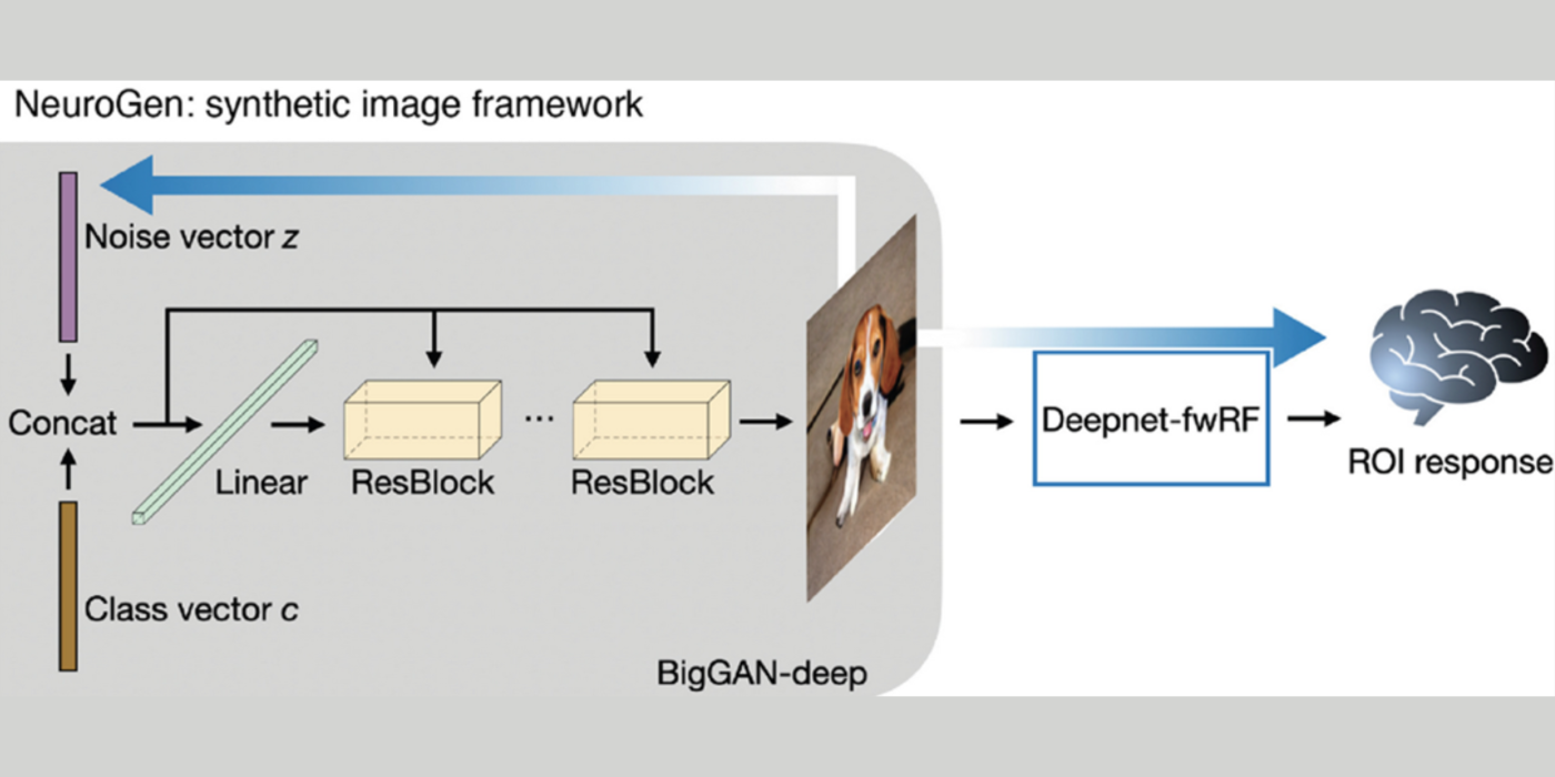
Zijin is a Ph.D. student in the Cornell University Department of Electrical and Computer Engineering. She received her bachelor’s degree in electrical engineering from Zhejiang University, China. Her research focuses on the intersection of machine learning and neuroscience. She is particularly interested in applying innovative machine learning algorithms to brain connectivity network analysis. Her project involves developing a noninvasive, spatially unconstrained and personalized method for neuromodulation, which involves creating deep neural networks for stimuli and brain activation patterns mapping. She hopes that manipulating brain connectivity networks will help alleviate symptoms or boost recovery after neurologic injury. 

View Bio
Lisa Iatckova
  • Ph.D. Candidate in Physiology, Biophysics, & Systems Biology

Lisa is a third-year student in the Weill Cornell Medicine Physiology, Biophysics and Systems Biology Graduate Program, and a recipient of a National Science Foundation Graduate Research Fellowship in bioengineering. After three years of studying economics in construction in her hometown in Russia, she immigrated to the U.S., and graduated with a B.A. in neurobiology from Hunter College in 2019. At Hunter, Lisa also conducted research in the Goldfarb lab, where she tricked brain cancer cells into expressing mutant sodium channels, and then poked them with electrodes to learn how the mutations affected voltage-dependent fast inactivation, a key property of the channel altered in disease phenotypes. Lisa’s strongest aspiration is to contribute to the development of brain-computer interfaces that improve quality of life for people with impaired physical and cognitive function (and maybe even augment natural brain capabilities of healthy people).  

Lab Focus

  • Human neuroimaging, mostly anatomical, functional and diffusion MRI.
  • Quantitative analysis of brain-behavior relationships.
  • Machine learning applied to human neuroimaging data.
  • Traumatic brain injury, multiple sclerosis, stroke and their recovery mechanisms.
  • Neuroimaging of psychedelics.
  • Sex and hormonal effects on the brain.

Lab Achievements

  • Editor’s Choice Collection 2019: awarded to the top six journal articles from that year’s publications, for the article: Kuceyeski A., Longitudinal increases in structural connectome segregation and functional connectome integration are associated with better recovery after mild TBI, Human Brain Mapping, 40 (15), p 4441-4456: 2019. 
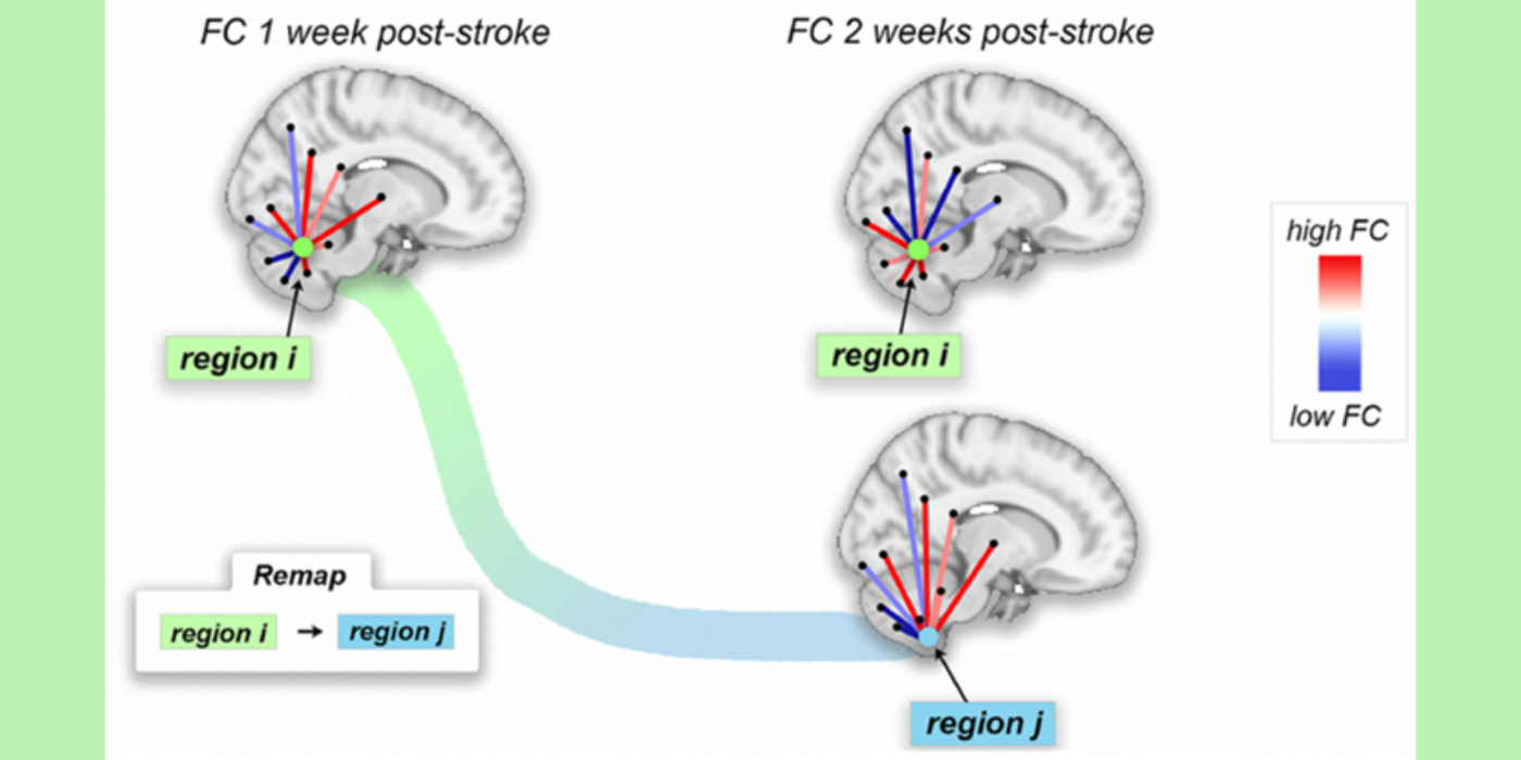
  • Organization for Human Brain Mapping 2021 Abstract Merit Award (top 1% of abstracts at the annual meeting), for the abstract, Functional Connectome Reorganization after Pontine Stroke is Associated with Better Motor Outcomes.
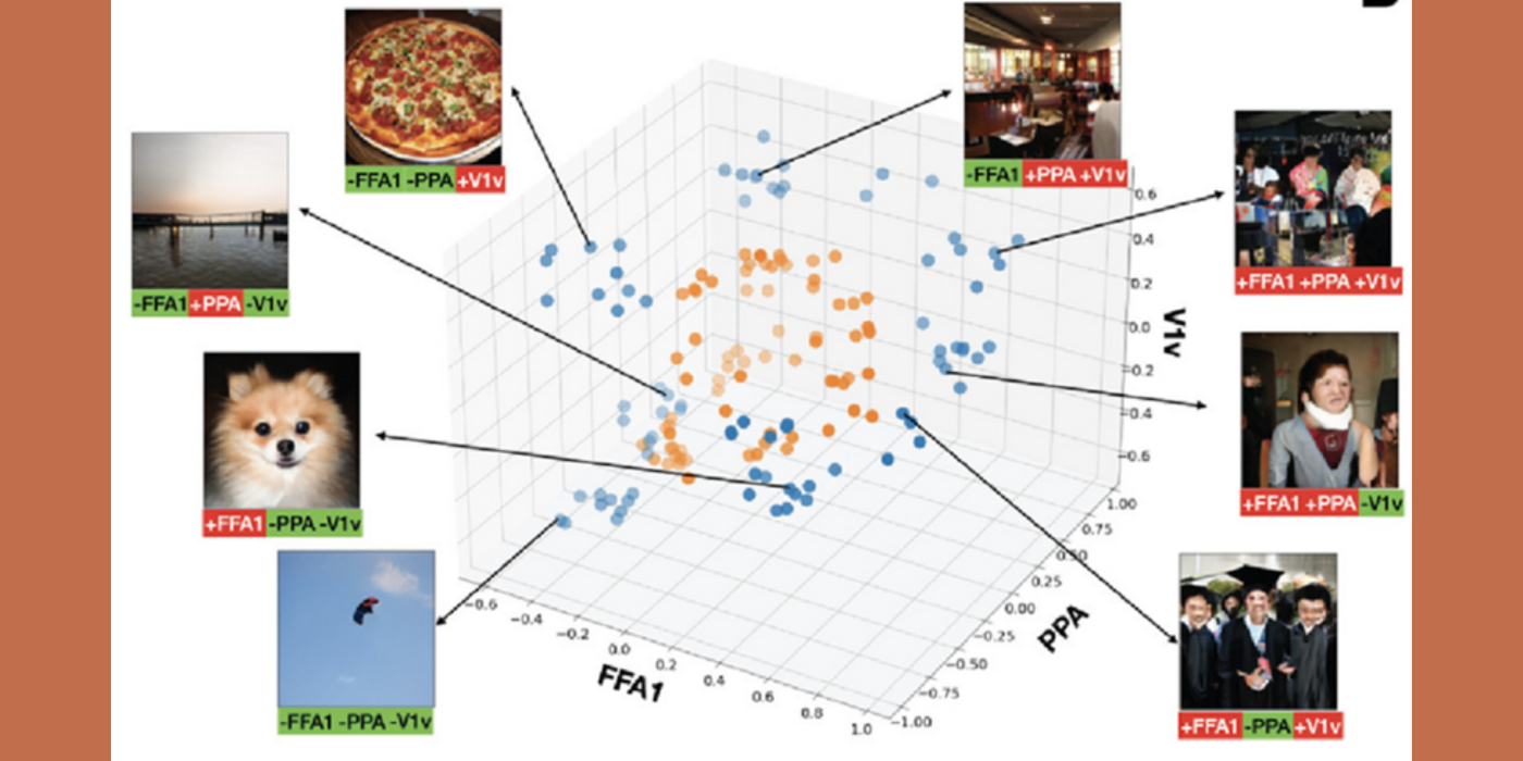
  • Organization for Human Brain Mapping 2021 Abstract Merit Award (top 1% of abstracts at the annual meeting), for the abstract, Identification and Synthesis of Preferred Images for Individual Regions in the Human Visual Cortex.

The year 2021 saw several publications from the CoCo lab in high-impact journals, including Nature Communications, NeuroImage, Human Brain Mapping, Cortex, Brain and Behavior, Science Advances, Current Opinion in Neurology, European Journal of Neurology and Frontiers in Neuroscience.

Collaborators

Kristen Dams-O'Connor, Ph.D.
Susan A. Gauthier, D.O.
Mert Sabuncu, Ph.D.

Research Projects

Award or grant: National Institutes of Health (NIH)/National Institute of Neurological Disorders and Stroke (NINDS),  1R01NS102646-01A1

Traumatic Brain Injury (TBI) is a...

Award or grant: National Institutes of Health/National Institute of Mental Health (NIH/NIMH): 1RF1MH123232

The human brain is an unimaginably complicated system of...

Award or grant: Leon Levy Foundation Fellowship

Understanding the organization of the white matter structural connectivity (SC) network in...

Award or grant:  National Institutes of Health/National Institute of Neurological Disorders and Stroke (NIH/NINNDS): R21 NS104634-01

Multiple Sclerosis (MS) damages white...

Motor recovery post-ischemic stroke relies on surviving brain networks' ability to compensate for damaged tissue. In rodent models, sensory and motor cortical representations remap onto intact tissue around lesion sites...

Award or grant: Cornell University MRI Facility pilot grant

Multiple sclerosis (MS) is one of the most common chronic inflammatory diseases wherein the body attacks protective tissue surrounding nerves. Women are more likely to develop MS compared to men with a...

Utilizing multi-modal neuroimaging techniques including fluorescence magnetic resonance imaging (fMRI), diffusion MRI (dMRI), and positron emission tomography (PET), the lab is applying new methods in network control theory to...

Neuromodulation (the localized manipulation of neuronal activity) holds therapeutic promise for neurological/neuropsychiatric diseases. There is currently an unmet need...

Weill Cornell Medicine
Department of Radiology
525 East 68th Street New York, NY 10065