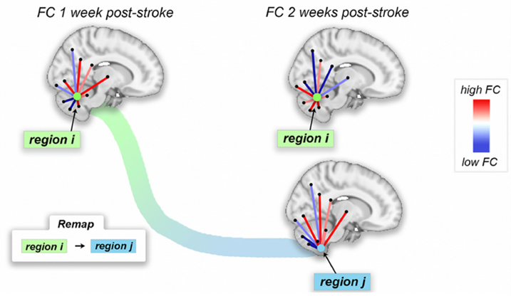
Motor recovery post-ischemic stroke relies on surviving brain networks' ability to compensate for damaged tissue. In rodent models, sensory and motor cortical representations remap onto intact tissue around lesion sites, but remapping to distal sites is seen. Resting-state functional connectivity (FC) analysis can unveil compensatory network adaptations in humans, but mechanisms/time course of motor recovery are unsure. Here we examine longitudinal FC in 23 first-episode ischemic pontine stroke patients via a graph-matching approach identifying recovery functional connectivity reorganization patterns. We quantified functional reorganization between intervals from one week to six months post-stroke, showing functional reorganization occurs most often in cerebellar/subcortical networks and areas with more structural and functional connectome disruption due to stroke. We show functional reorganization correlates with the extent of motor recovery in early- to late-subacute phases, and those with greater baseline motor impairment show more extensive early subacute functional reorganization (from one to two weeks post-stroke), which correlates with better motor recovery at six months. This approach quantifies recovery-relevant, whole-brain functional connectivity network reorganization post-stroke. Above: regions that remap.


