Associated Lab Members

Dr. Tracy Butler is a neurologist/neuroscientist with clinical subspecialty training in behavioral neurology and epilepsy and research fellowship training in functional and structural neuroimaging. She is the medical director of the Brain Health Imaging Institute (BHII) where she oversees subject assessment and clinical trials of therapies and neuroimage biomarkers of aging and neurodegeneration. Her research uses multimodal positron emission tomorgraphy (PET) and magnetic resonance imaging (MRI) neuroimaging and complementary methods to better understand the biological basis of neuropsychiatric disorders including Alzheimer’s disease, traumatic brain injury, and as normal aging, focusing on pathophysiologic overlap among these conditions such as hormonal dysregulation, neuroinflammation and brain toxic protein (tau and amyloid) accumulation.

In 2020, Dr. Farnia Feiz joined the Quantitative Neuroimaging Laboratory (QNL) as clinical research manager. She helps with patient recruitment, medical data review, and regulatory operations of the QNL’s National Institutes of Health-funded study. Prior to arriving at Weill Cornell Medicine, Dr. Feiz worked on numerous studies in neuroradiology and neurodegenerative diseases. She holds an M.D. from Shiraz University of Medical Sciences and an M.P.H. from New York University.

Sarah is a research coordinator in the Tracy Butler lab for The LUCINDA trial. She is a graduate of Hunter College. She previously worked at Mount Sinai on a project investigating the basis of Chronic Fatigue Syndrome. She is pursuing a master’s degree in nutrition.

Tom Maloney is a highly experienced project and data manager for complex clinical research studies of brain function. He has deep expertise in developing database structures and tools and facilities for smooth, flexible capture, organization and retrieval of very large, very dense, highly diverse study data from multiple research and clinical domains. His background ranges from event-related electroencephalogram (EEG) potentials, sleep regulation, cognitive performance, chronobiology and neuropsychological testing to functional neuroimaging. He has planned and managed research and clinical operations and data migrations at Stony Brook University, the Veterans Administration, University of California, Brown University, Brookhaven National Laboratory, Mount Sinai School of Medicine, and, since 2019, Weill Cornell Medicine. He is project manager for the LUCINDA Trial and Data Manager for the Brain Health Imaging Institute.
Xiuyuan (Hugh) Wang has been working with Dr. Butler since 2011, first at New York University and now at Weill Cornell. He received his bachelor's degree in biomedical engineering from Shanghai University, and a master's degree from City College of New York. He is an image analyst in the lab working on biomedical signal and image processing.
Dr. Tracy Butler is a neurologist/neuroscientist with clinical subspecialty training in behavioral neurology and epilepsy and research fellowship training in functional and structural neuroimaging. She is the medical director of the Brain Health Imaging Institute (BHII) where she oversees subject assessment and clinical trials of therapies and neuroimage biomarkers of aging and neurodegeneration. Her research uses multimodal positron emission tomography (PET) and magnetic resonance (MR) neuroimaging and complementary methods to better understand the biological basis of neuropsychiatric disorders including Alzheimer’s disease (AD), traumatic brain injury (TBI), and normal aging.
Dr. Butler’s clinical work is limited to serving as the neurology and neuropsychiatry consultant to NewYork-Presbyterian Behavioral Health Center, diagnosing and treating hospitalized patients with severe psychiatric diseases who can sometimes fall between the cracks of psychiatry and neurology.
We use PET with novel and Food and Drug Administration (FDA)-approved radiotracers, functional and structural MRI, and complementary neuroimaging and other techniques to improve understanding and treatment of human brain disorders including AD, TBI, epilepsy, depression, and normal aging. We focus on pathophysiologic overlap among these conditions such as hormonal dysregulation, neuroinflammation, and brain toxic protein accumulation and clearance.
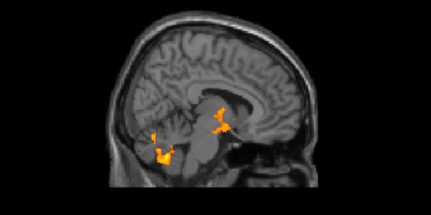
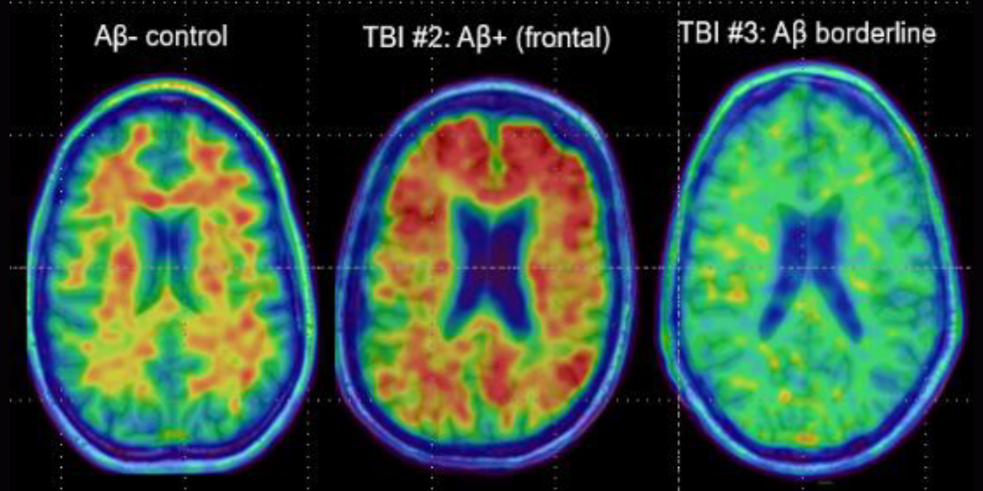
Traumatic Brain Injury (TBI) is a leading cause of death and disability worldwide and a risk factor for later development of Alzheimer's disease (AD). TBI causes increased axonal production and rapid brain deposition of amyloid, a pathologic hallmark of AD. Persistence of amyloid...
LUCINDA is a clinical trial to determine whether Leuprolide (...
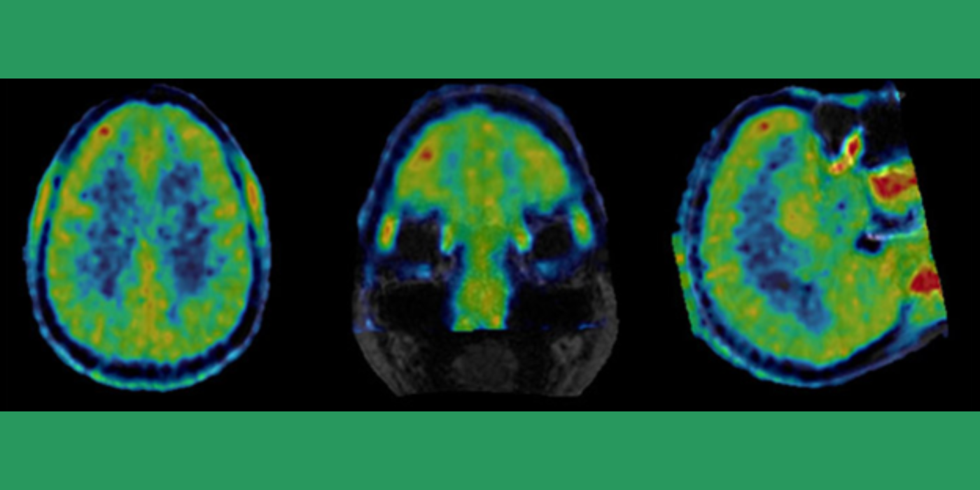
Inflammation is a vital, complex process by which certain cells or tissues identified by the immune system as abnormal (e.g., foreign, damaged or dead) can be repaired or removed to facilitate continued survival of remaining cells and the organism. Excessive or dysregulated...
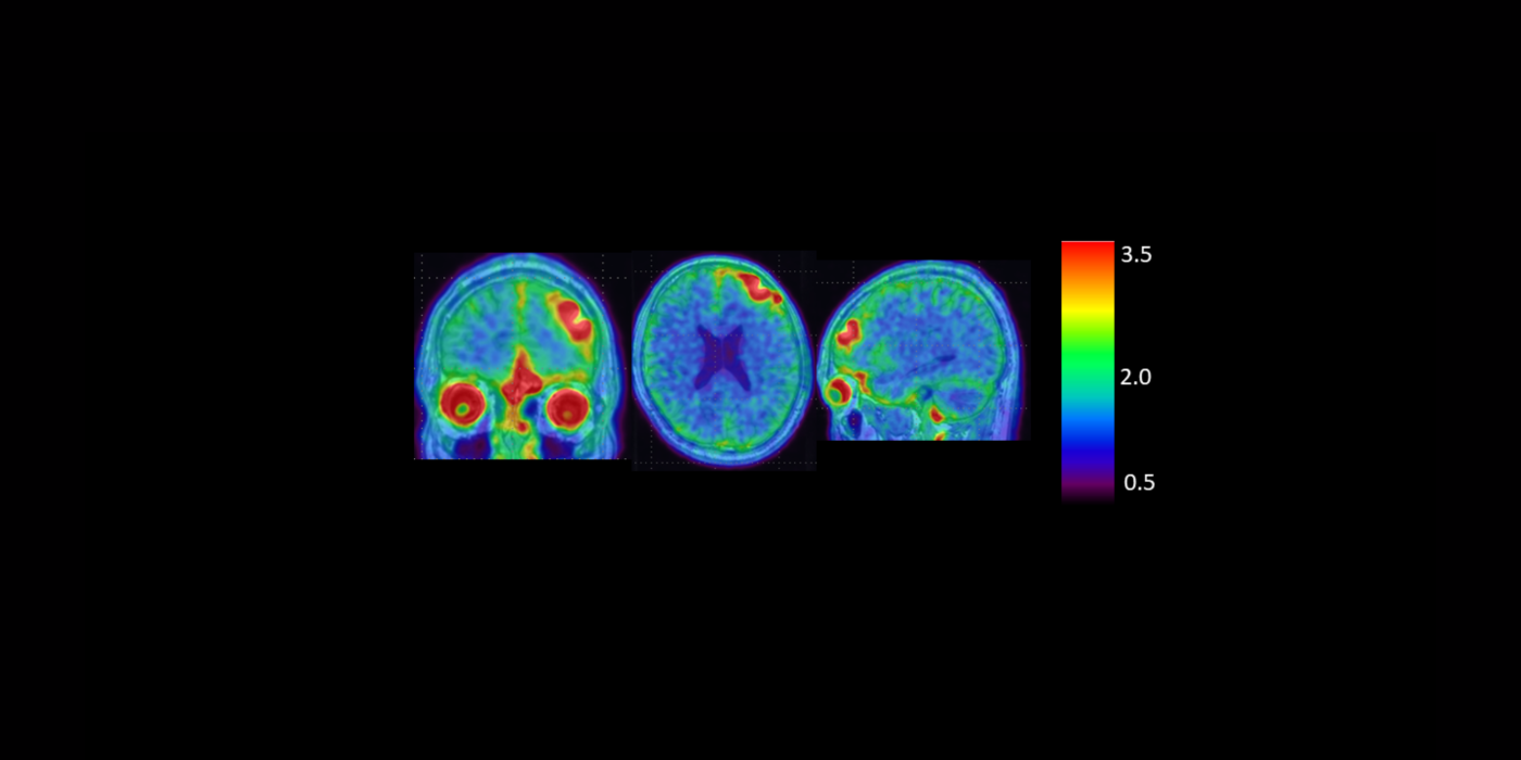
This pilot project will assess positron emission tomography (PET) and magnetic resonance imaging (MRI) biomarkers of neurodegeneration in female victims of intimate partner violence (IPV) with and without TBI. The goal is to...
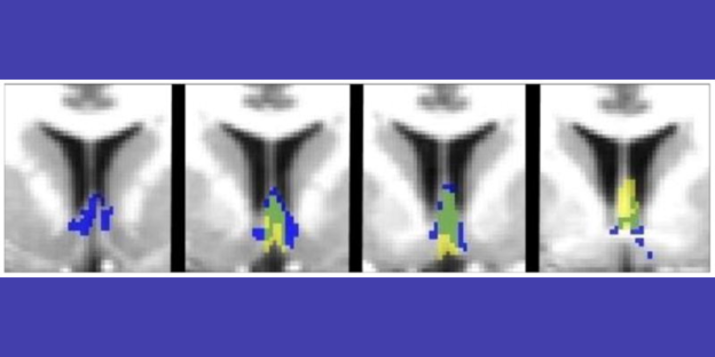
Septal nuclei, located in the anterior basal forebrain, exert strong control over...