The lab’s studies began with general characterizations of brain damage, and were first to characterize neocortical atrophy, positron emission tomography (PET)-imaged glucose metabolism, and white matter lesions as clinical features of Alzheimer’s dementia (AD). Subsequent lab studies expanded understanding of the anatomy of memory; developed early AD predictive hippocampal formation tests; introduced cerebrospinal fluid (CSF) biomarkers that conferred pathological specificity; and examined clinical and genetic risks for AD. The lab is now working on a new PET-based radioligand technology to evaluate CSF dynamics. The de Leon team’s current clinical research also targets vascular factors that contribute to AD lesions, oral bacterial dysbiosis leading to brain inflammation and amyloidosis, traumatic brain injury as a risk factor for AD, sleep abnormalities and tau pathology, and the mechanistic role of inflammation in the failed removal of waste product from brain. The team has developed one of the world’s largest repositories of matched blood and CSF with magnetic resonance imaging (MRI) and PET imaging. Dr. de Leon has received many awards, and has been well funded by the National Institutes of Health (NIH). He was in the top 5% of extramural NIH grantees for 25 years.
Associated Lab Members

Mony J. de Leon, Ed.D., is director of the Brain Health Imaging Institute (BHII). Trained in gerontology and neuroscience, his research focuses on the clinical detection of brain changes underlying cognitive dysfunction, and the characterization of mechanisms contributing to misfolded brain proteins and tissue damage in aging and Alzheimer’s disease (AD). His team’s post-mortem validated biomarker discoveries of hippocampal atrophy and brain glucose metabolism deficits predicting clinical outcomes have contributed to the current standard diagnostic assessment of AD.
Mohammad Khalafi, M.D., a graduate of Tabriz University of Medical Sciences, reviews medical data and contributes to ongoing research projects at the Brain Health Imaging Institute. Before joining Weill Cornell Medicine, Dr. Khalafi researched neuroradiology and neurodegenerative diseases.
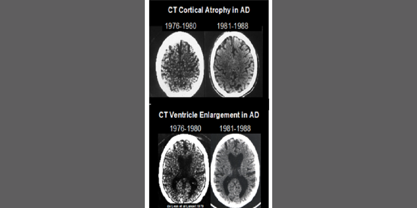
De Leon lab early studies were first to show that cortical atrophy in dementia patients can be quantitatively evaluated using computed tomography (CT), and can serve, after ventriculomegaly, as the second imaging biomarker for age-related neurodegenerative disease. In the 1980s, this helped lead to the inclusion of structural imaging findings in diagnostic workups for dementia.
Selected papers: de Leon, M.J., Correlations between CT changes and behavioral deficits in senile dementia, Lancet, 859-860: Oct. 20, 1979; de Leon, M.J., et al., Longitudinal CT studies of ventricular change in Alzheimer's disease, AJNR, 10, 371-376: 1989.
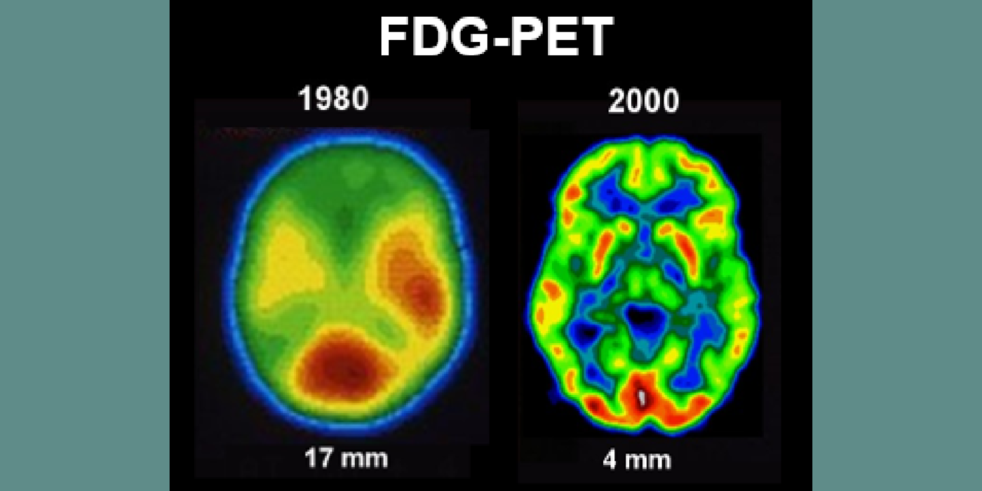
In a collaboration between the Brookhaven National Labs, New York University, and the University of Pennsylvania, Dr. de Leon reported the first fluorodeoxyglucose (FDG)-PET AD studies. These studies identified widespread neocortical metabolism reductions in dementia patients. The team’s later studies unearthed early metabolic reductions in the entorhinal cortex and hippocampus of normal aging subjects at risk for future cognitive change. These longitudinal FDG-PET study subjects, followed to post-mortem, demonstrated that the hippocampal formation changes were accurately predictive of the post-mortem diagnosis of AD.
Selected papers: Ferris, S.H., et al., Positron emission tomography in the study of aging and senile dementia, Neurobiol. Aging, 1, 127-131: 1980; de Leon, M.J., et al., Prediction of cognitive decline in normal elderly subjects with 2-[18F]fluoro-2-deoxy-D-glucose/positron-emission tomography (FDG/PET), Proc. Natl. Acad. Sci., 98, 10966-10971: 2001. Mosconi, L., et al., FDG-PET changes in brain glucose metabolism from normal cognition to pathologically verified Alzheimer's disease, Eur. J. Nucl. Med., 36(5), 811-822: 2008.
The team’s anatomical studies identified age-related periventricular white matter lesions on CT and (later) MRI scans. The team developed the first white-matter rating scale and showed these CT lesions were clinically associated with hypertension and, at pathology, with microvascular pathology. This work flourished in the MRI era.
Selected papers: George, A.E., et al., Leukoencephalopathy in normal and pathologic aging 1. CT of brain lucencies, AJNR, 7:561-6: 1986; George, A.E., et al., Leukoencephalopathy in normal and pathologic aging: 2. MRI and brain lucencies, AJNR, 7:567-70: 1986; de Leon, M.J., George, Altered patterns of positron emission tomography glucose metabolism in Alzheimer patients with microvascular white matter disease, Am. J. Physiol., 3, 52-53): 1988; Marcus, D.L., et al., Altered glucose metabolism in microvessels from patients with Alzheimer's disease, Ann. Neurol., 26, 91-94: 1989.
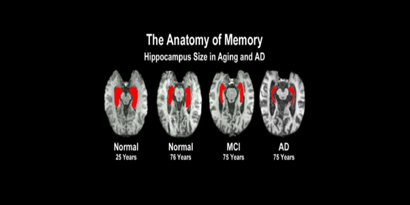
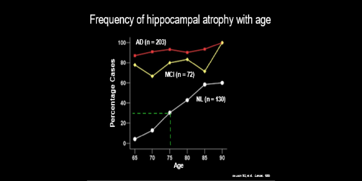
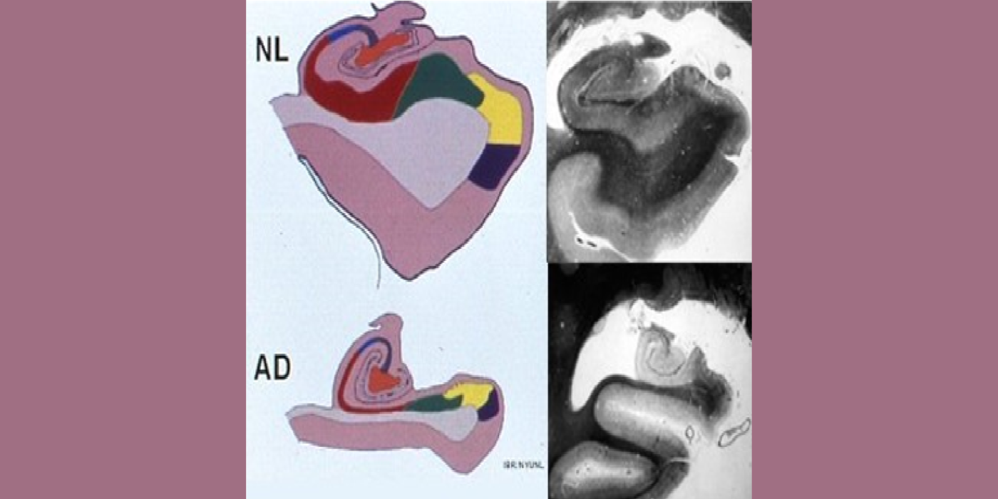
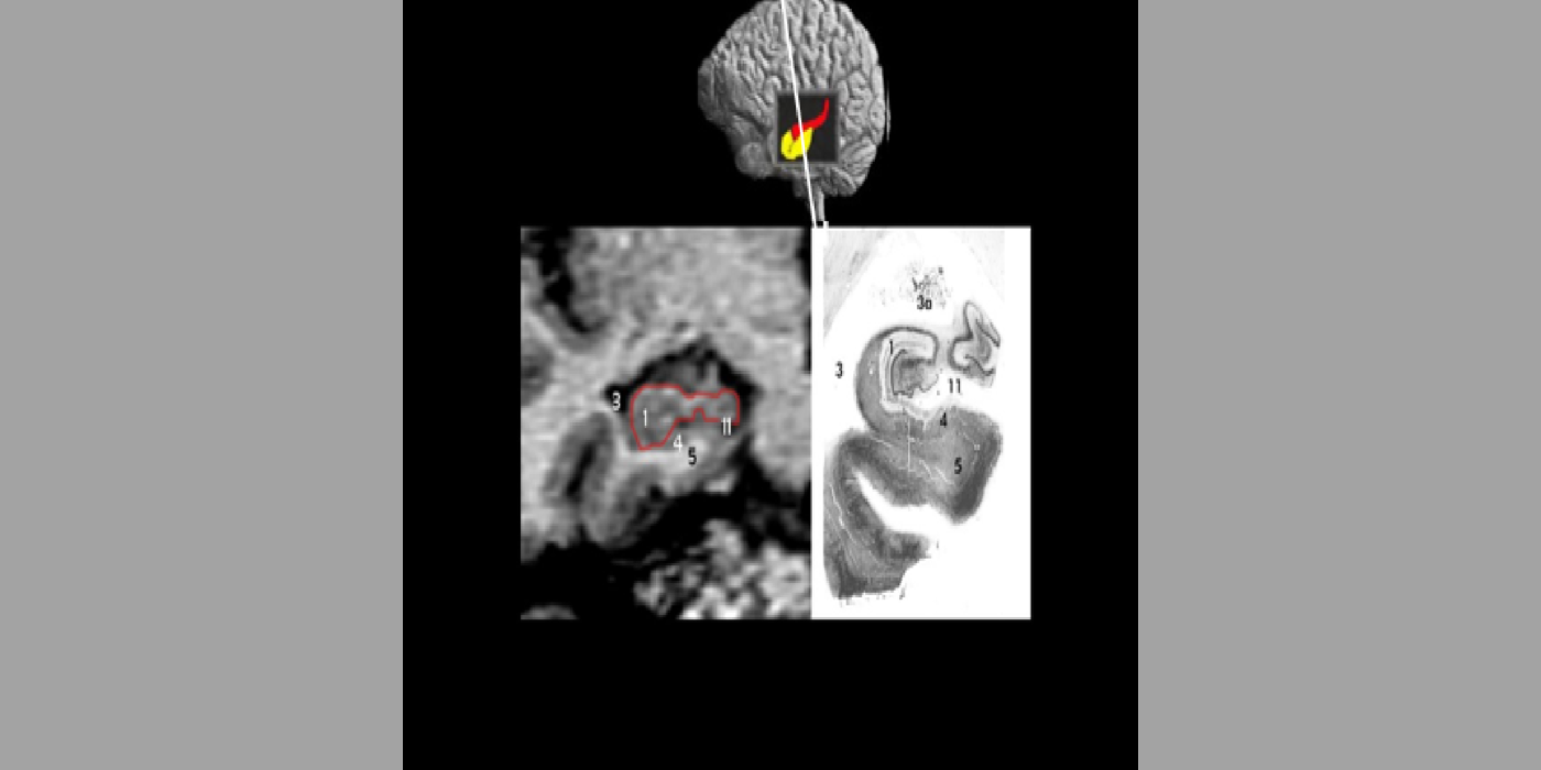
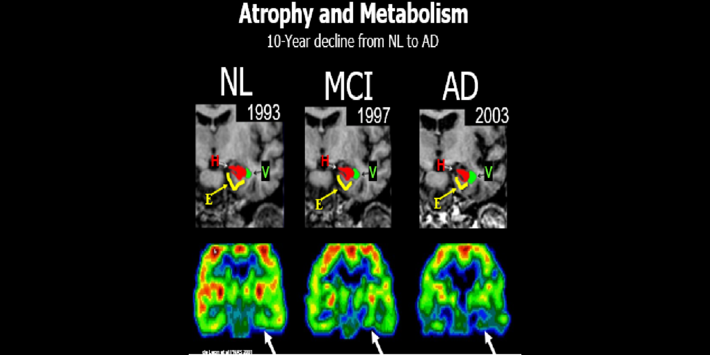
With evolving instrumentation, combined with novel image acquisition and longitudinal experimental designs, smaller targets—including atrophic changes of the hippocampus and the entorhinal cortex— allow the identification of subjects with normal cognition. But a fortuitous turning point emerged in de Leon lab ventricular anatomy studies. Using negative CT angulation to image the axis of the temporal horn of the lateral ventricle, the lab observed excess atrophic changes in the hippocampal region in subjects with AD (1988). These studies also characterized the prevalence of hippocampal atrophy in aging (30% of normal subjects with atrophy by age 75) and >80% of subjects atrophic with dementia.
Subsequent hippocampal imaging led to the first MCI-AD prediction study (1989) and the first normal-cognition-to-MCI prediction study using MRI (high resolution (HR)) and FDG-PET (2001). This benefitted from Dr. de Leon’s collaboration with psychiatrist Barry Reisberg (the first description of MCI in 1982), and pathologist Henrik Wisniewski (the first anatomic and pathologic validations of the hippocampus in subjects with the de Leon team’s in vivo imaging). This led to the awarding, to Dr. de Leon, of The Pioneer in Imaging Award, Alois Alzheimer Centennial, Tubingen Germany, 2006. (See Carousel Images 3-6.)
Selected papers: de Leon, M.J., et al., Abnormal cortisol response in Alzheimer's disease linked to hippocampal atrophy, Lancet, 2(8607), 391-392: 1988; de Leon, M.J., et al., Early marker for Alzheimer's disease: The atrophic hippocampus, Lancet, (672-673) Sept. 16, 1989; Rusinek, H., et al., Regional brain atrophy rate predicts future cognitive decline: 6-year longitudinal MR imaging study of normal aging, Radiology; 229(3):691-6: Dec. 2003; Bobinski M., et al., The histological validation of post mortem magnetic resonance imaging-determined hippocampal volume in Alzheimer's disease, Neurosci; 95: 721-5, 2000 ; Bobinski, M., et al., MRI of entorhinal cortex in mild Alzheimer's disease, Lancet 353, 38-40: 1999.
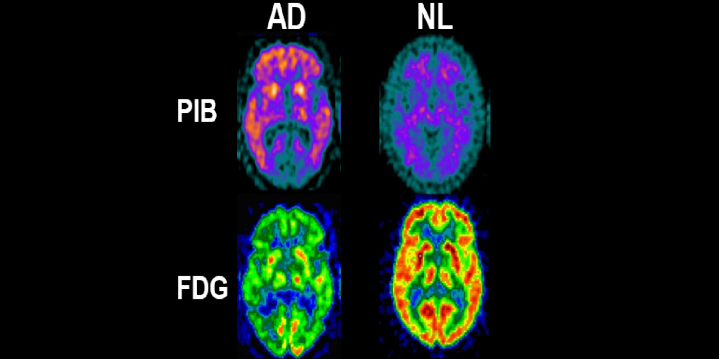
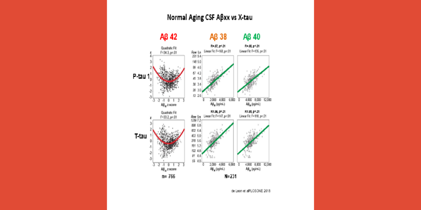
A major turning point in the AD biomarker field was first introduced by the availability of CSF biomarkers of Aβ and tauopathy and later, PET imaging biomarkers for neuropathology. These studies were revolutionary as they conferred disease specificity into the clinical exam. Plasma tests are now available. The BHII routinely recruits subjects with family AD histories, ApoE genotype, hypertension, impaired sleep, joint replacement surgery, brain trauma, and periodontal bacterial infections, and COVID-19. The BHII uses imaging, CSF, and plasma AD biomarkers in longitudinal designs to study disease progression and therapeutic interventions.
Selected papers: de Leon M.J., et al., CSF clearance in Alzheimer Disease measured with dynamic PET, J Nuc Med; 58(9):1471-1476: Sept. 2017; Glodzik, L., et al., Different Relationship Between Systolic Blood Pressure and Cerebral Perfusion in Subjects with and without hypertension, Hypertension, 73(1):197-205: Jan. 2019; Osorio,.R., et al., The interaction between sleep-disordered breathing and apolipoprotein E genotype on cerebrospinal fluid biomarkers for Alzheimer's disease in cognitively normal elderly individuals, Neurobiol. Aging, ISSN 0197-4580, 2013; Kamer, A.R., et al., Periodontal dysbiosis associates with reduced CSF Aβ42 in cognitively normal elderly, Alzheimers Demen.; 13(1):e12172: Apr. 12, 2021.
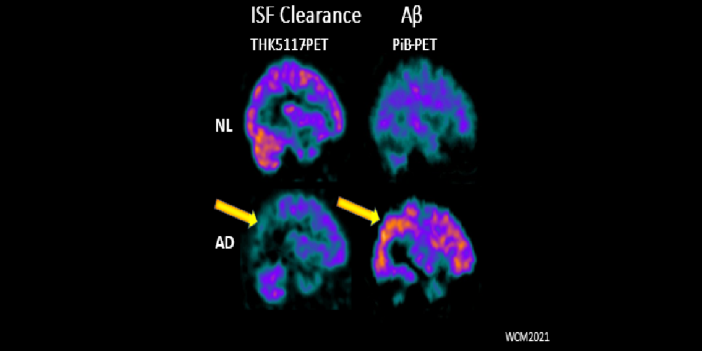
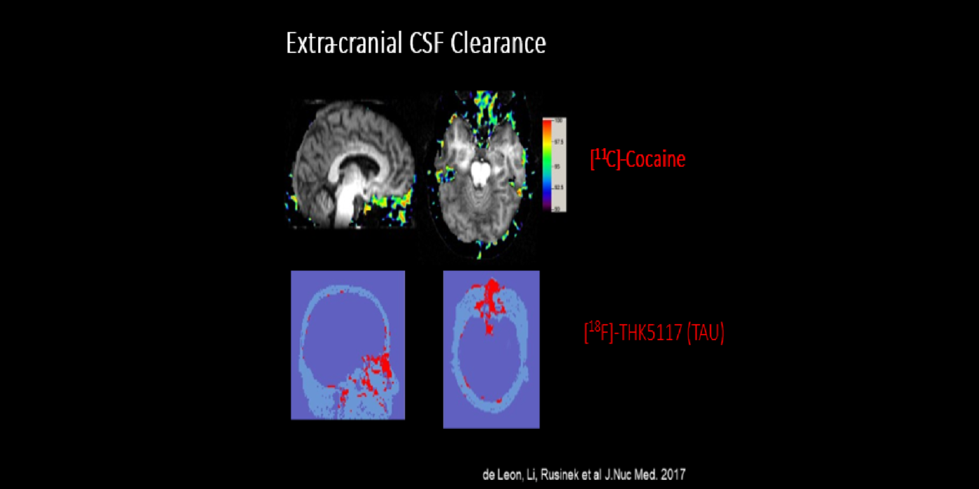
It has been hypothesized amyloid beta (Aβ) clearance is reduced in AD, potentially a causal factor for its fibrillization and accumulation. We tested the hypothesis that reduced CSF clearance was associated with amyloid accumulation using 11C-Pittsburgh compound B (PiB). We observed CSF clearance, measured from the lateral ventricle using radiotracer 18F-THK5117, was reduced in AD, and associated with the magnitude of amyloid accumulation. Little is known about the extracranial egress of CSF from the human brain. By examining the extracranial compartment with two radiotracers, we identified a nasal egress pathway through the cribriform plate into the superior nasal turbinates.
Selected papers: Tarasoff-Conway, J.M., et al., CSF clearance in Alzheimer Disease measured with dynamic PET, J. Nucl. Med.,58(9):1471-1476: Sept. 2017; Mehta, N.H., Front. Physiol., 12:769948: Jan. 4, 2022.
Tracy Butler, M.D.
Yi Li, M.D., Ph.D.
Anna Nordvig, M.D.
David Pisapia, M.D.
Yi Wang, Ph.D.
Award or Grant: National Institutes of Health (NIH)/National Institute on Aging (NIA) 1RF1 AG057570 (09/15/17-03/31/22).
This project is designed to develop and test a positron emission tomography (PET) based ventricular...
Awards or Grants: 2R44AG044860-04, National Institutes of Health (NIH) (03/01/17-02/28/23)
The main objective of this collaborative project is to assess the microRNA-based assay performance for the detection of mild cognitive...
Awards or Grants: R56 AG058913, National Institutes of Health/National Institute of Aging (NIH/NIA) (09/15/18-08/31/22)
This positron emission tomography (PET) project examines...
Awards or Grants: R01 AG013616, National Institutes of Health/National Institute of Aging (NIH/NIA) (09/01/91-03/31/17)
This project, renewed multiple times, followed an aging cohort with blood and cerebrospinal fluid (CSF) sampling and...
Awards or Grants: AG022374, National Institutes of Health/National Institute of Aging (09/30/10-05/31/18)
The combined use of magnetic resonance imaging (MRI) hippocampal atrophy and cerebrospinal fluid (CSF)...
Awards or Grants: AG012101, National Institutes of Health/National Institute of Aging (NIH/NIA), 09/15/10-05/31/18
Testing cerebral blood flow using arterial spin labeling (ASL)-magnetic resonance imaging (MRI) with inhaled carbon dioxide (CO2) as a predictor of...