The McIntire Lab uses state-of-the-art lipidomic techniques including imaging mass spectrometry to interrogate the specific and critical changes to the homeostasis in the lipidome in mouse models, induced pluripotent stem cells (iPSC), and human biospecimens. Mounting evidence indicates that dysregulation of the lipidome is strongly implicated in neurodegenerative diseases such as Alzheimer's disease (AD) as well as aging. Genetic studies have identified multiple genes in lipid metabolic pathways associated with AD risk and for amelioration of behavioral deficits in mouse models of AD. Most notably, a variant of apolipoprotein E (ApoEε4), a cholesterol transporter in the brain, is the strongest genetic risk factor for late-onset AD. Lipidomic data confirm lipid dyshomeostasis in autopsy brain, cerebral spinal fluid, human plasma, and animal models pointing to the potential for specific pathway(s) in lipid metabolism to underlie multiple AD disease mechanisms. However, there has not yet been a system-wide analysis able to synthesize these mounting data.
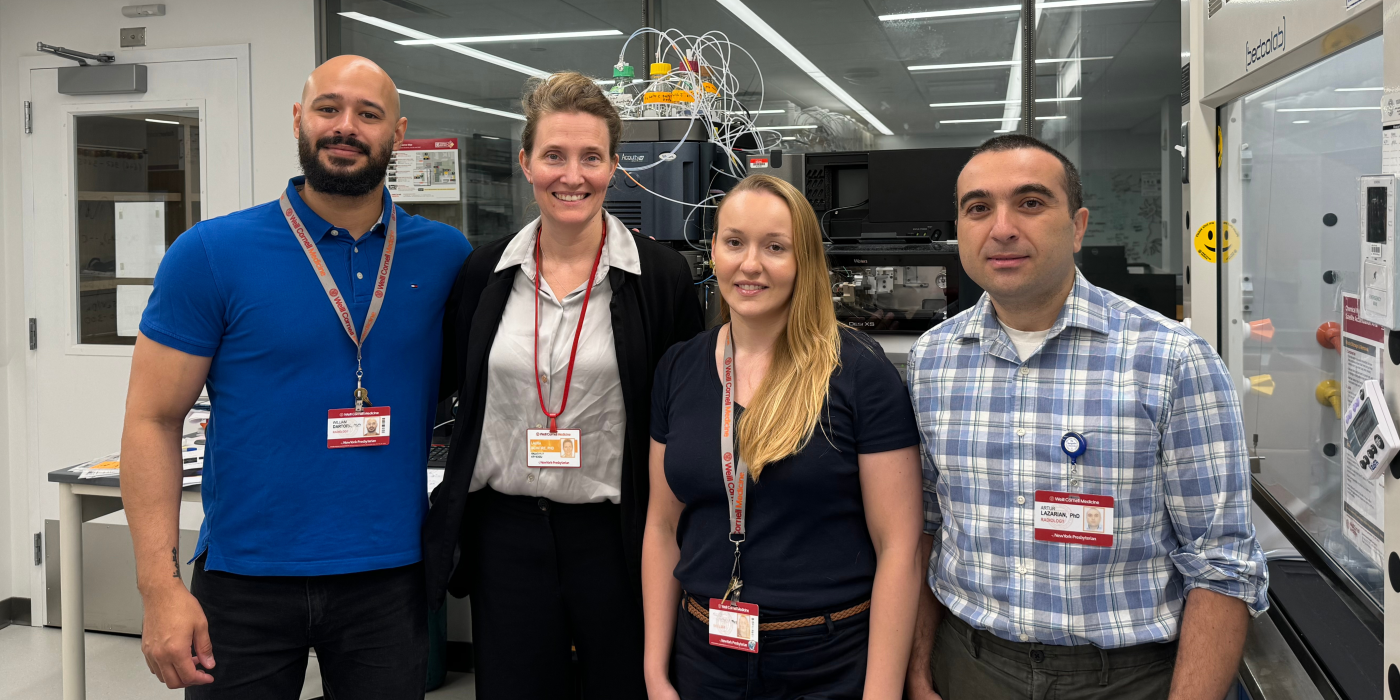
Associated Lab Members

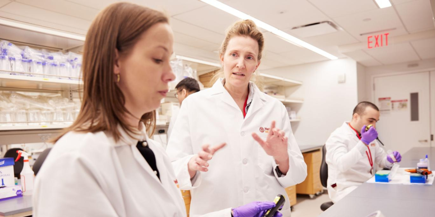
Dr. Laura Beth McIntire, Ph.D., is director of the Lipidomics and Biomarker Lab at the Brain Health Imaging Institute (BHII), and an assistant professor of pharmacology, in the department of radiology. Her work focuses on the contribution of lipid dyshomeostasis to Alzheimer’s disease (AD) with a focus on phosphoinositide metabolism and acyl chain remodeling. Dr. McIntire is an expert in cell biology, cellular imaging and animal models of AD with expertise in animal behavior and the generation of novel mouse strains. She has expertise in lipidomics, imaging mass spectrometry, and system-wide analyses of the lipid interactome which have led to novel insights into lipid deficits in AD progression.

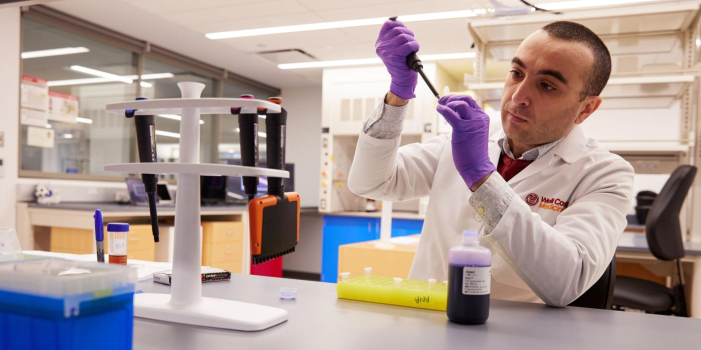
Dr. Artur Lazarian, Ph.D., has extensive experience with mass spectrometry, molecular biology and biochemistry. His recent work has focused on desorption electrospray ionization (DESI) imaging mass spectrometry using the Waters Synapt G2-Si. Additionally, during his previous post-doc position at the Max-Planck Institute for Polymer Research, he extensively used the Waters Synapt G2-Si. In the past two years, he has expanded his expertise to include molecular modeling using Schrodinger software.
Optogenetic control of phosphoinositides as ameliorative target in AD mouse model
Synj1 reduction ameliorated AD-associated synaptic and behavioral deficits in a mouse model of AD, lending evidence that Synj1 may be a promising therapeutic target. To further investigate this premise, the lab uses optogenetics to temporally and spatially regulate phosphoinositide lipid synthesis in mouse brain for validation of phosphoinositides as critical players in resistance and resiliency to AD. The lab has created a light-induced dimerization system with a phosphoinositide kinase catalytic domain (CD) PIP5K2A-CD fused to cryptochrome 2 (CRY2) (PIP5K2A-CD-CRY2-mCherry) and the membrane targeted CRY2-binding domain (CIBN) CIBN-CAAX which resulted in increased phosphatidylinositol 4,5-bisphosphate (PIP2) at the plasma membrane. The lab found amelioration of behavioral deficits in both Novel Object Recognition and Contextual Fear Conditioning. To determine differential phospohinositide levels in neurodegenerative diseases, the lab compares human plasma, cerebrospinal fluid (CSF) and brain tissue from patients with AD and traumatic brain injury in targeted lipidomics studies.
Functional interrogation of gene network including Synaptojanin1 and GWAS identified hits BIN1 and PICALM in iPSC- derived neurons.
Further supporting the premise of the critical nature of lipid dyshomeostasis in AD, results from a candidate based screen conducted in my lab for mitigation of amyloid-beta (Aβ)-triggered synapse loss resulted in identification of synaptojanin 1 (Synj1), phospholipase D (PLD) 1/2, multiple phospholipase C (PLC) isoforms, phosphoinositide phosphatases and the genome wide association studies (GWAS) hit Bridging Integrator 1(BIN1). BIN1 is a predicted to interact with Synj1, phosphatidylinositol binding clathrin assembly protein (PICALM) and factors important for regulating endocytosis. Due to trisomy of Synj1 on chromosome 21 in Down syndrome (DS) and several phenotypic commonalities to both AD and DS such as enlarged endosomes and dendritic spine alterations, targeting Synj1 has potential for unmet therapeutic need in DS as well as AD.
Identification of dysregulated lipid pathways for biomarker and therapeutic target discovery
To test the hypothesis that manipulation of brain lipid content alters brain lipidome and cognitive function in mouse models of AD, the lab proposes to genetically alter the synthetic enzyme, Acyl CoA Synthetase 6 (Acsl6), a key mediator of polyunsaturated fatty acid enrichment in the brain especially docosahexaenoic acid (DHA) across multiple lipid classes, in mouse models of AD. Lipid content will be determined in brain of mouse models of AD (APPsw and TRAPOE4) using imaging mass spectrometry for integration of a publically available lipid brain atlas.
We have identified deficits in acyl chain remodeling that are associated with disease progression in AD and can be harnessed for amelioration of behavioral deficits in a mouse model of disease. We have also:
The McIntire lab is studying changes in the brain lipidome using targeted liquid chromatography/mass spectrometry (LC/MS) and untargeted imaging mass spectrometry (IMS) resulting in a system-wide, publically available lipid map. Data from the lab show distinct regional distribution of acyl chain...
This project involved the generation of a novel mouse strain that ameliorated phosphoinositide depletion and behavioral deficits despite the continued accumulation of amyloid.
Using optogenetics to temporally and spatially regulate phosphoinositide lipid synthesis in the mouse brain, the McIntire lab has shown amelioration of phosphoinositide dysregulation and behavioral benefit in a mouse model of Alzheimer's disease (AD).
Screening for phosphoinositide-modifying enzymes that can prevent amyloid-induced synaptic loss in mouse embryonic stem cell-derived neurons.