Award or Grant: R01 AG079326-01
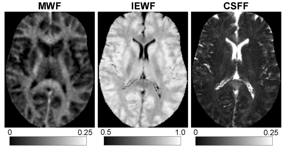
The team’s objective is to develop a new imaging method for quantitative mapping of parenchymal cerebrospinal fluid fraction (CSFF). The team’s preliminary data showed CSFF increased in Alzheimer’s disease (AD) and was associated with cerebrospinal fluid (CSF) bulk flow reduction and decreased ventricular CSF clearance. More importantly, increased CSFF correlated with brain amyloid (studied using 11C-PiB positron emission tomography (PET)) and tau (studied using 18 fluorodeoxyglucose (F)-MK6240 PET). The team’s postmortem data also showed an increased CSFF and perivascular space load in the AD brain. The team proposes to complete, over five years, a two-year longitudinal study designed to test, in preclinical AD, the hypothesis that CSFF is associated with ventricular CSF clearance (measured by dynamic 18F-MK6240 PET) and bulk CSF flow (measured by phase-contrast magnetic resonance imaging (MRI)), and that increased CSFF is predictive of future amyloid lesions (amyloid-PET), brain tau load (18F-MK6240 PET), and cognitive decline (memory composite) in early-stage AD. This project also includes a post-mortem study to verify glymphatic pathology in different disease stages.
The team’s pilot data showed a significant correlation between CSFF, CSF clearance function, and brain amyloid and tau deposition, indicating that the proposed study could confirm the utility of CSFF in quantifying the dynamic relationship between glymphatic clearance and amyloid beta (Aβ) and tau accumulation in AD. A successful outcome of this project will further our understanding of the pivotal role of glymphatic clearance in brain disorders including AD, and potentially identify much-needed new therapeutic targets.


