Coronary Ischemia, when to intervene? Coronary Artery Disease impacts more than 18 million worldwide and was responsible for ~350,000 deaths in 2019. In the case of coronary ischemia, where coronary stenosis results in reduced blood flow to the myocardium, one challenge is determining when disease is severe enough to warrant intervention (e.g., stent, bypass, angioplasty). Today, the gold standard for this determination is a hemodynamic measure known as fractional flow research (FFR), obtained by measuring pressure drop across a stenosis using a minimally invasive pressure measurement catheter. When the pressure drop is large (FFR<0.8), interventions are warranted. When it is small (FFR>0.8), typically patients can be treated with traditional therapies until the disease becomes more severe.
The need for non-invasive measures of ischemic severity. While FFR remains the gold standard to prognosticate coronary ischemia, there is a desire for effective measures that do not require minimally invasive catheterization. Today, one promising approach involves the use of 3D modeling and subsequent computational fluid dynamic analysis of coronary computed tomography angiography (CCTA). In this approach, 3D models of the coronary vessels are segmented from CCTA images and the pressure drop across the coronary stenosis is calculated based on the geometry of the vessel. This allows for an effective estimation of FFR without the need for direct measurement.
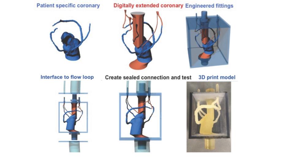
Materials matter: Soft Materials and 3D printing. While fluid dynamic estimations of FFR provide a compelling non-invasive measure of stenosis severity, these calculations have limitations. In particular, they typically assume that the walls of the vessels are entirely rigid. Calculations that consider the tissue-like properties vessel are challenging and require significant computational power. In order to study the importance of vessel mechanics on these hemodynamic measures, our lab is using multi-material 3D printing to produce realistic coronary models with tissue-like mechanical properties. Recent advances in 3D printing allow for fabrication of coronary models with complex mechanical properties that vary spatially within the model. Based on high-quality CCTA images, this allows us to create models that accurately recapitulate complex and tortuous vessel geometries, but model the complex and varying mechanical properties of real coronary disease. By deploying these models in simulated flow loops with realistic conditions, we can study the way that FFR and other hemodynamic measurements are impacted by mechanical properties.


