The long-term objective of this project is to improve the ability to monitor inflammatory and neurodegenerative processes in multiple sclerosis (MS) for effective diagnosis and treatment. The specific goal: to establish the time course of magnetic susceptibility of lesions as a sensitive probe of demyelination and iron content associated with inflammatory processes. Currently, gadolinium enhancement in conventional magnetic resonance imaging (MRI) is widely used to map inflammation caused by blood brain barrier (BBB) disruption during MS lesion formation. However, there is still inflammation in the brain after the BBB heals, and currently there is no method in clinical practice to detect this inflammation behind the BBB. Our preliminary data demonstrate that the time course of lesion magnetic susceptibility as measured by quantitative susceptibility mapping (QSM) can detect lesion activity after the resolution of gadolinium enhancement. Accordingly, this proposed research will focus on establishing the existence—and gaining a basic understanding—of the time course of MS lesion susceptibility by carrying out three specific aims.
Aim 1: Improve QSM scanning robustness and processing accuracy for reliable longitudinal measurement of susceptibility time course. The lab hypothesizes that QSM can be improved to a high reproducibility and accuracy of 0.01ppm (parts per million, susceptibility unit). Aim 2: Define the MS lesion magnetic susceptibility time course through prospective longitudinal study of the susceptibility of lesions at all ages, with spatiotemporal correlation of gadolinium enhancement measured by conventional MRI. The lab hypothesizes that lesion susceptibility changes dynamically over time, even after inflammatory activity, as measured by conventional MRI, has subsided. Aim 3: Define the immunohistochemical correlates of MS lesion magnetic susceptibility using autopsied brain specimens from MS patients and controls. The lab hypothesizes that the QSM signal of an MS lesion correlates in part with microglia/macrophages activation (i.e., inflammatory activity) and that the QSM value of a fully demyelinated lesion is determined by iron.
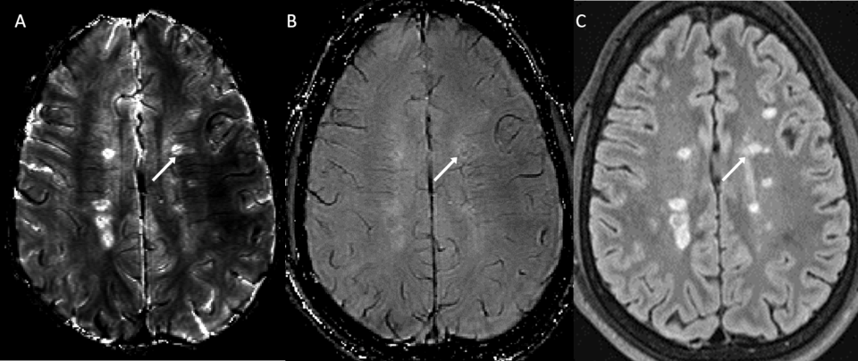
(Full Caption) Male, 38, with multiple sclerosis. His central vein sign was judged positive on susceptibility relaxation optimization (SRO) (A), negative on susceptibility weighted imaging (SWI) for a fluid-attenuated inversion recovery (B) and hyperintense lesion (C) (arrow). The central vein is highly conspicuous on SRO due to the enhanced contrast between its hypointensity and the surrounding hyperintense lesion signal. The central vein exhibits poor contrast on SWI. J. Neuroimag.: 2021.


