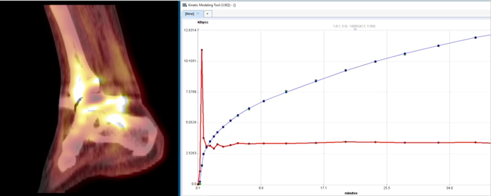
Bone blood flow (BBF) and turnover may be altered in many disease states such as osteoarthritis (OA), bone marrow lesions, and trauma or fracture. Dr. Dyke’s group pioneered the use of pharmacokinetic magnetic resonance imaging (MRI) and positron emission tomography (PET) modeling of various agents to measure BBF and turnover in disease progression of OA, and following injury. These techniques are being used to assess bone healing following total ankle replacement at increasing times post-surgery.
Associated Departments: Departments of Radiology and Orthopedic Surgery, Weill Cornell Medicine and Hospital for Special Services
Related Publications: (1) Dyke J.P., et al., Imaging of Bone Perfusion and Metabolism in Subjects Undergoing Total Ankle Arthroplasty Using 18F-Fluoride Positron Emission Tomography, FAI: 2019;40:1351-57. (2) Aaron R.K., et al., Subchondral Bone Circulation in Osteoarthritis of the Human Knee, Osteoarthr. Cartil: 2018;26:940-944. (3) Dyke J.P., et al., Characterization of Bone Perfusion by Dynamic Contrast-Enhanced Magnetic Resonance Imaging and Positron Emission Tomography in the Dunkin-Hartley Guinea Pig Model of Advanced Osteoarthritis, J. Orthop. Res.: 2015;33:366-72. (4) Dyke J.P., Regional analysis of femoral head perfusion following displaced fractures of the femoral neck, J. Magn. Reson. Imaging: 2015;41:550-554.


