Award or Grant: R01CA219964, National Cancer Institute (NCI) (02/01/18 – 01/31/23).
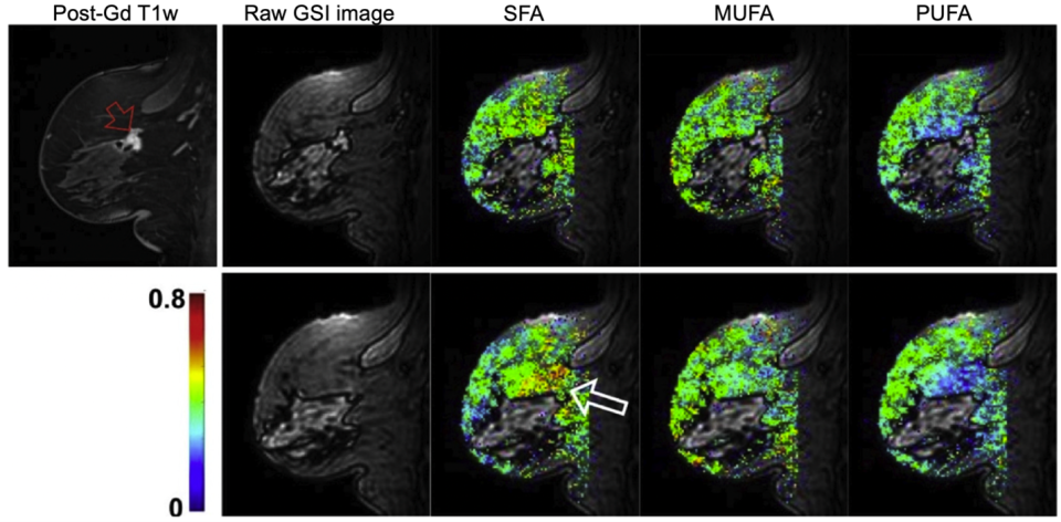
Description: The role of breast fatty acid composition in breast cancer development, in conjunction with breast density and body mass index, has not been fully investigated. The central hypothesis of the Kim lab is that its recently developed gradient-echo spectroscopic imaging method can be used to investigate the association of the spatial distribution of saturated fatty acids in breast adipose tissue with the presence of malignant lesions. This study is conducted on patients undergoing diagnostic breast magnetic resonance imaging (MRI) exams or MRI-guided core biopsy exams.
Project Summary: The role of fat in breast cancer development and growth has been studied extensively using body mass index (a measure of whole-body fat), and dietary fat intake, in many epidemiological studies. However, there is a paucity of studies, on an individual level, to assess the role of fat itself in breast cancer due to the lack of a fast non-invasive measurement method. Since breast fibroglandular cells are surrounded by breast fat cells, the characteristics of breast fat may be tightly related to breast cancer development, as supported by recent studies showing that a majority of breast cancers develop at the interface between fibroglandular tissue and adipose tissue. But it is not trivial to study the role of breast fat, as noted, due to the lack of a fast non-invasive method sensitive enough to measure key features of breast fat, i.e., fat types.
The Kim lab has developed a rapid MRI method, gradient-echo spectroscopic imaging (GSI), to measure fatty acid composition during clinical breast MRI exams. GSI can provide maps of saturated and unsaturated fats in breast adipose tissue without requiring tissue biopsy. Our pilot study found postmenopausal women with aggressive invasive ductal carcinoma have a significantly higher percentage of saturated fat in their breast adipose tissue than postmenopausal women with only benign lesions. The goal of this study is to determine the role of saturated fatty acid in breast cancer development/growth. The lab will use GSI for non-invasive in vivo measurement of saturated fat in the breast adipose tissue of postmenopausal women undergoing diagnostic breast MRI exams (Aims 1 and 2) or MRI-guided biopsy scans (Aim 3). Our central hypotheses: (i) breast-saturated fatty acid fraction measured by GSI is associated with malignant lesions in the breast; (ii) breast-saturated fatty acid fraction correlates positively with inflammation in breast adipose tissue—which may lead to an increase in adipocyte estrogen production.
This study will evaluate whether breast saturated fat is an independent risk factor for breast cancer and whether it can provide additional diagnostic information to current clinical diagnostic exams. Our imaging measure of breast saturated fat can be used to assess the efficacy of any intervention to reduce cancer-related inflammation in breast adipose tissue and to investigate the possible role of fatty acid composition in the prevention and clinical management of breast cancer.


