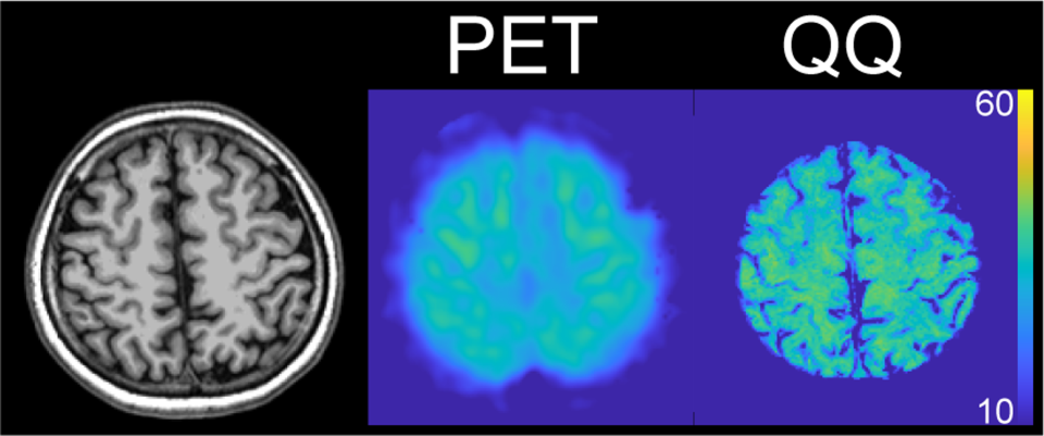
The objective of this proposed research is to develop a noninvasive, easily accessible and widely usable imaging method to map the cerebral metabolic rate of oxygen consumption (CMRO2). Oxidative metabolism is the main source of energy for proper human brain function. Consequently, brain tissue is highly susceptible to damage associated with oxygen deficiency, including hypoxia in Alzheimer’s disease and multiple sclerosis, and ischemia in stroke. Quantitative mapping of CMRO2 (qmCMRO2) would be very valuable for studying various brain functions and for evaluating these neurologic disorders. Magnetic resonance imaging (MRI) is widely distributed, offering the potential to overcome the 15O positron emission tomography (PET) availability problem. Cerebral blood flow (CBF) can be mapped using spatially selective radiofrequency (RF) to generate arterial spin labeling (ASL) or using a contrast agent to label spins. qmCMRO2 requires estimating dH concentration ([dH]) from the MRI signal. Three qmCMRO2 approaches have been developed for extracting [dH] from MRI, all utilizing only the magnitude signal that unfortunately has a complex dependence on [dH]. Consequently, current MRI-based qmCMRO2 suffers from poor sensitivity. It is cumbersome to perform in research and difficult to use in clinics. Furthermore, none of these MRI-based qmCMRO2 methods have been validated by the current reference standard, 15O PET. The lab proposes to utilize the often-discarded MRI phase signal quantitative susceptibility mapping (QSM) pioneered by the Wang lab.
In this R21 project, the lab will establish a Bayesian qmCMRO2 that is challenge-free by optimally utilizing both phase and magnitude data from gradient echo (GRE) MRI, managing uncertainties in biological priors, and validating the developed challenge-free MRI-based qmCMRO2 using 15O PET. The lab will achieve this through two specific aims. Aim 1: Develop fast challenge-free qmCMRO2 using GRE and ASL MRI. Aim 2: Validate challenge-free MRI-based qmCMRO2 using 15O PET on a PET/MR hybrid scanner. The lab’s experience and preliminary data give it confidence that it will very likely succeed with this proposed project. In a timely fashion, the project will lead to an easily and widely used tool for quantitative mapping of CMRO2, which can be routinely used on all the extensively distributed 3 tesla (T) MRI scanners to study brain functions and neurological diseases.


