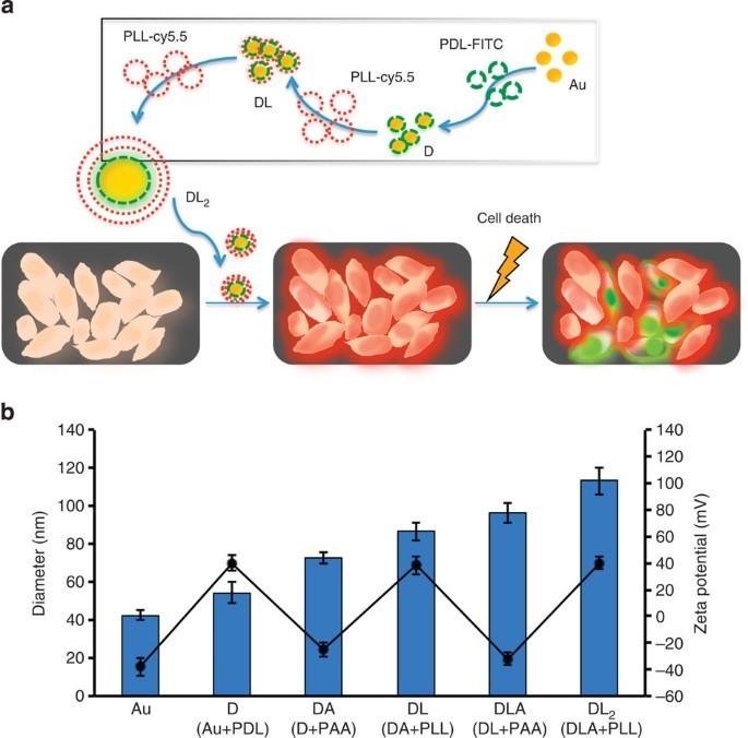
Using layer-by-layer nanoplatform (LbLN) technology, Seung Koo Lee invented a state-of-the-art cell fate tracker (DL2) that showed the vital status of cells, whether they were alive or dead, in a Nature Communications paper with the above title. In that paper, he noted that tracing of cell viability was key for determining cell therapy safety and viability. Dr. Lee therefore created a nanoparticle-based cell tracker to image cell fate from live to dead. This particle was fabricated from two types of optically quenched polyelectrolytes, one a life indicator and the other a death indicator, through electrostatic interactions. Once incubated with cells, the bifunctional nanoprobes were taken up efficiently and produced a color by intracellular proteolysis. This indicated the cells were healthy. Depending on the number of coated layers, the signal could persist for several replication cycles. When they began dying, a second color reflected that new state. In their abstract, Dr. Lee and the paper’s senior author, Dr. Ching Tung, called this a “chameleon-like cell tracker,” which could distinguish live from apoptotic and necrotic cells “instantly and definitively.”
Dr. Lee and colleagues have since gone on to:
- Demonstrate, for a gene delivery system, that a small interfering RNA (siRNA)-formulated LbLN could silence target-gene expression for more than a three-week period in breast cancer xenograft models (Lee et al., Adv. Funct. Mater.: 2013).
- Apply miRNA-708 (Ramchandani et al., Mol. Cancer Ther.: 2019) or Bcl-xL siRNA and KLA mitochondria targeting peptide (Chen et al., Mol. Ther.-Oncolytics: 2021) to LBLN, achieving significant therapeutic effects for breast cancer and pancreatic cancer.
- Develop novel fluorescence probes for multiple diagnostic applications that can be used in gene therapy, drug delivery and enzymatic molecular imaging (Lee et al., Macromol. Biosci.: 2019).


