Awards or Grants: R01 CA160620, National Cancer Institute (NCI), 2/1/2019-1/31/2024
Description: Assessment of cancer treatment requires an effective non-invasive method of measuring both the vascular and cellular changes induced by therapies. The Kim team’s underlying hypothesis is that a single dynamic contrast-enhanced (DCE) magnetic resonance imaging (MRI) measurement, using the active contrast encoding (ACE) MRI method, can provide a fast and quantitative means to assess both anti-angiogenic and cytotoxic responses to therapy. This study will establish an innovative method of assessing chemotherapy that will significantly improve drug discovery and patient management in many cancers including breast cancer.
Project Summary: Despite recent development of new approaches to therapy, breast cancer remains the second leading cause of cancer death in women. Treatment with anti-angiogenic drugs, combined with conventional cytotoxic drugs or immunotherapy, is a promising means of treating aggressive cancer. Anti-angiogenic drugs are thought to temporarily normalize abnormal vasculature and paradoxically increase blood flow and hence delivery of drug and effector immune cells to tumors. But a substantial proportion of patients do not respond to this combination therapy. It is unclear whether this is due to failure of the anti-angiogenic to normalize the vasculature, or failure of the cytotoxic drugs or immune cells to kill cancer cells. It is thus necessary to assess both blood flow and cell death to elucidate mechanism, and optimize the combination treatments. In this study the lab investigates one MRI acquisition and analysis, allowing assessment of both.
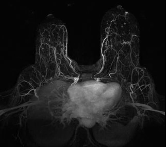
DCE MRI has been widely used as an important part of most clinical MRI exams for cancer diagnosis, possessing great potential as a single MRI method to estimate both perfusion parameters (such as flow, F, vascular volume fraction, vp, and vascular permeability-surface area product, PS) and cellular parameters (such as interstitial volume fraction, ve, and intracellular water lifetime, ti). Recently, we developed a novel data acquisition method, ACE-MRI, which measures dynamic data together with pre-contrast T1 and B1 that are critical for measurement of perfusion and cellular parameters. ACE-MRI is also implemented with a fast 3D imaging method to acquire high-spatial and high-temporal resolution data using a 3D golden-angle ultra-short echo-time (UTE) sequence and an image reconstruction method to combine both compressed sensing and parallel imaging, also known as GRASP (Golden-angle RAdial Sparsity and Parallel). In this study, the Kim team will further develop ACE-MRI to accurately estimate contrast agent concentration in vascular plasma and tissue using a direct blood sampling method (Aim 1), and to assess the association of ti with tumor metabolic rate and treatment response in comparison with 18F-fluorodeoxyglucose (FDG)-positron emission tomography (PET) and pathology (Aim 2). Overall, the ACE-MRI parameters will be used to assess treatment response and metastatic potential (Aim 3). This study is conducted with murine and human breast cancer models at a 7 tesla (T) small animal MRI scanner. However, the methods developed can be easily translated to clinical applications, as they are used on an ultra short time echo sequence readily available on most clinical scanners.


