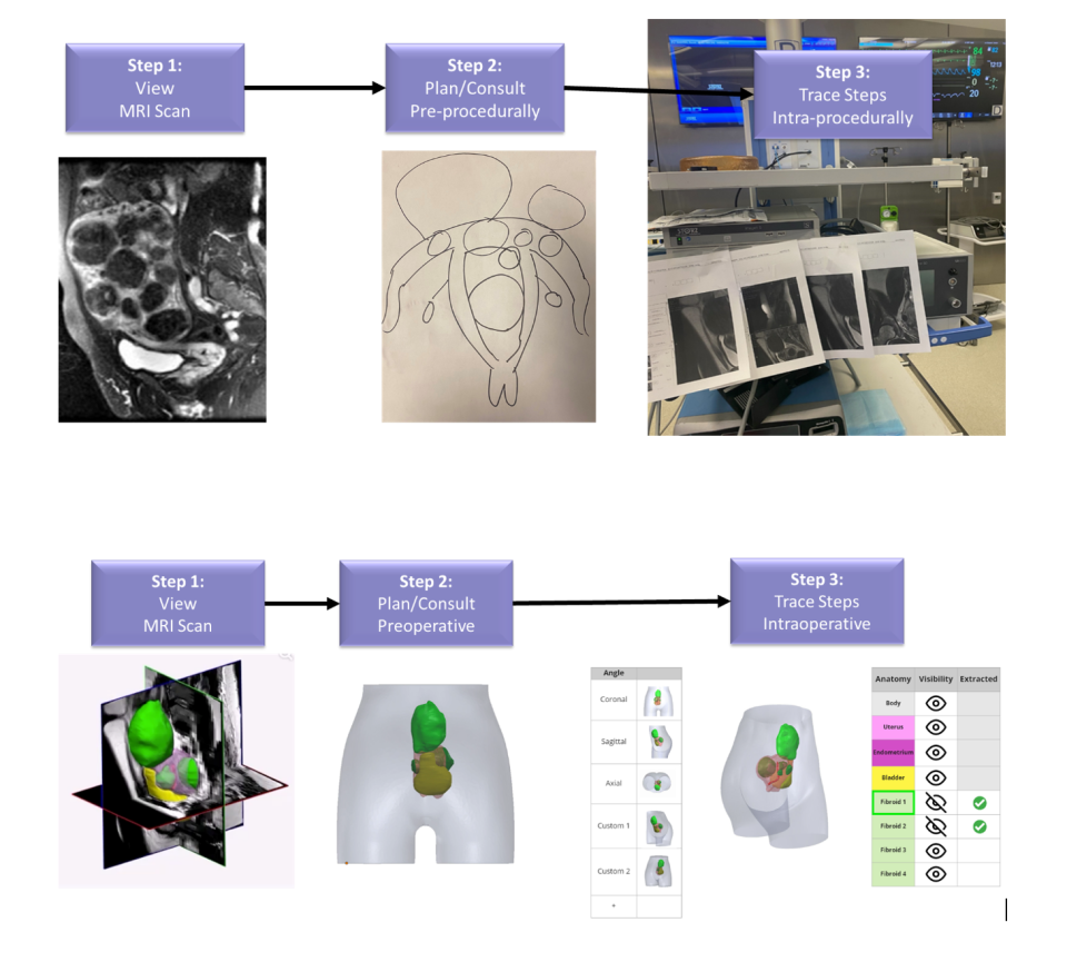
Most benign tumors are uterine fibroids in women, especially African American women. Today, doctors counsel women using flat magnetic resonance imaging (MRI) images, but fibroids can grow on multiple uterine planes, and 2D imaging does not accurately allow optimized planning and intraoperative guidance. Our technology brings MRI into the modern age by leveraging deep learning algorithms to generate 3D renderings. Physicians can interact with these before procedures to improve diagnosis and path planning, and during procedures to optimize adherence to that plan with fewer complications and faster procedural durations. The lab has institutional review board (IRB) approval for a clinical trial to assess the usefulness of this technology for patient counseling and minimally invasive surgery. Goal: characterize the improved benefits offered by 3D visualization on both 2D screens and mixed reality headsets, as opposed to standard MRI viewers.
Full Caption: Workflows of today and tomorrow. (Top) Current workflow. (1) Radiologist views 2D MRI scans. (2) Patient advised with the help of paper images or 2D MRI slices. (3) Guidance: memorizing and displaying fibroid positions on 2D screens/printing slices. (Bot.) Next-Gen workflow. (1) MRI scan auto-segmented by deep learning model. Radiologist views 3D scans embedded on 2D slices. (2) Patient advised via 3D rendering of segmented fibroids. (3) Guidance: voice-command, interactive, mixed reality fibroid removal.


