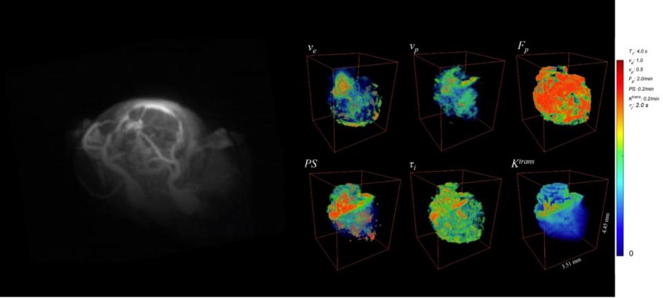
Assessment of cancer treatment requires an effective non-invasive method of measuring both the vascular and cellular changes induced by therapies. Our central hypotheses is that dynamic contrast-enhanced (DCE) magnetic resonance imaging (MRI) measurement, using the active contrast encoding (ACE) MRI method, can provide a fast and quantitative means to assess both anti-angiogenic and cytotoxic responses to therapy. To enhance the spatial and temporal resolution for 3D DCE-MRI experiments, we have developed a 3D ultra-short echo time (UTE) Golden-angle Radial Sparse Parallel (GRASP) MRI method. This figure displays a representative example of the GL261 mouse tumor model. The left panel shows a 3D volume rendering of the raw image, while the right panel exhibits 3D pharmacokinetic parameter maps