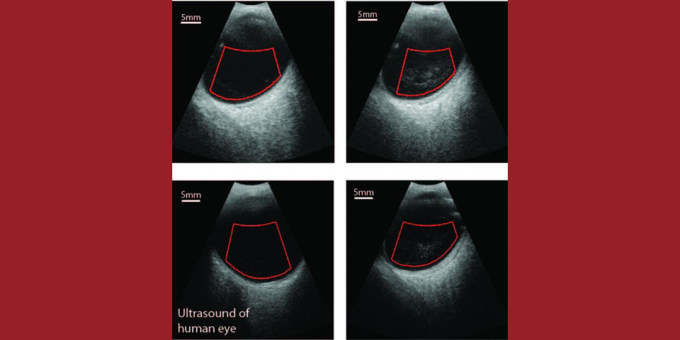
The goal of this translational project is to add quantitative ultrasound (QUS) capabilities to a clinical ophthalmic ultrasound system to characterize vitreous inhomogeneity (i.e., clinically significant vitreous floaters referred to as Vision Degrading Myodesopsia) as it relates to states of health and disease. Age-related changes due to collagen cross-linking and aggregation with liquefaction create inhomogeneities that appear non-uniformly throughout the vitreous body. For patients with myopia, these processes occur earlier in life, when vitreo-retinal adhesion is still strong, and destabilize the vitreous body before adhesion to the retina is weakened, resulting in various conditions that impact vision. The ability to depict vitreo-retinal organization will offer unique early-stage detection, and assessment, of vitreo-retinal disease in patients with myopia at risk for retinal detachment and vitreo-maculopathies resulting from traction. Currently, no diagnostic method is available to make data-based decisions related to the changes taking place in the vitreous body before blinding pathologies have developed. The impending epidemic of myopia has created an urgent clinical need for technologies offering objective and sensitive means for early detection of macromolecular changes and structural precursors in the vitreous body directly related to vitreo-retinal diseases. A diagnostic tool capable of quantitatively characterizing the entire vitreous body would assist in developing less invasive and affordable treatments for early intervention, such as pharmacologic vitreolysis or laser therapy, and for identifying patients in need of more aggressive interventions with vitrectomy. In collaboration with Quantel Medical, we will incorporate a new 3D probe and QUS capabilities into a state-of-the-art, 20-MHz annular-array-based clinical ophthalmic ultrasound system. The early stages of the project will emphasize device enhancements to obtain necessary raw ultrasound data, followed by age-normal and myopic patient data collection and development of QUS algorithms to characterize the vitreous body in 2D. The later stages of the project will focus on development and integration of a probe capable of volumetric acquisition, further patient data collection, and QUS classification methods that take advantage of the new volumetric data. The final system will permit quantitative characterization of the vitreous body and allow for data-based treatment decisions of vitreo-retinal diseases. While this investigation will focus on myopia, the resultant technology and approach will have the potential for application in many different clinical settings.


