Tracy Butler Laboratory

- Associate Professor of Neurology in Radiology
Dr. Tracy Butler is a neurologist/neuroscientist with clinical subspecialty training in behavioral neurology and epilepsy and research fellowship training in functional and structural neuroimaging. She is the medical director of the Brain Health Imaging Institute (BHII) where she oversees subject assessment and clinical trials of therapies and neuroimage biomarkers of aging and neurodegeneration. Her research uses multimodal positron emission tomorgraphy (PET) and magnetic resonance imaging (MRI) neuroimaging and complementary methods to better understand the biological basis of neuropsychiatric disorders including Alzheimer’s disease, traumatic brain injury, and as normal aging, focusing on pathophysiologic overlap among these conditions such as hormonal dysregulation, neuroinflammation and brain toxic protein (tau and amyloid) accumulation.
- Research Associate in Radiology
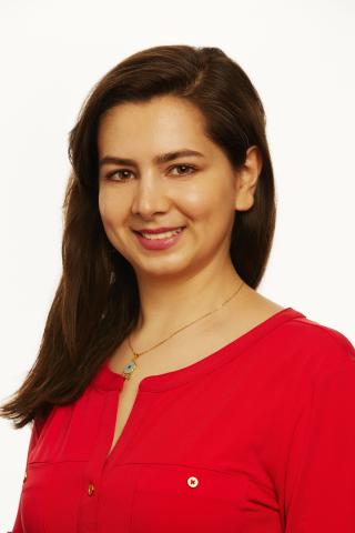
In 2020, Dr. Farnia Feiz joined the Quantitative Neuroimaging Laboratory (QNL) as clinical research manager. She helps with patient recruitment, medical data review, and regulatory operations of the QNL’s National Institutes of Health-funded study. Prior to arriving at Weill Cornell Medicine, Dr. Feiz worked on numerous studies in neuroradiology and neurodegenerative diseases. She holds an M.D. from Shiraz University of Medical Sciences and an M.P.H. from New York University.

- Research Coordinator in Radiology
Sarah is a research coordinator in the Tracy Butler lab for The LUCINDA trial. She is a graduate of Hunter College. She previously worked at Mount Sinai on a project investigating the basis of Chronic Fatigue Syndrome. She is pursuing a master’s degree in nutrition.

- Senior Research Associate in Radiology
Tom Maloney is a highly experienced project and data manager for complex clinical research studies of brain function. He has deep expertise in developing database structures and tools and facilities for smooth, flexible capture, organization and retrieval of very large, very dense, highly diverse study data from multiple research and clinical domains. His background ranges from event-related electroencephalogram (EEG) potentials, sleep regulation, cognitive performance, chronobiology and neuropsychological testing to functional neuroimaging. He has planned and managed research and clinical operations and data migrations at Stony Brook University, the Veterans Administration, University of California, Brown University, Brookhaven National Laboratory, Mount Sinai School of Medicine, and, since 2019, Weill Cornell Medicine. He is project manager for the LUCINDA Trial and Data Manager for the Brain Health Imaging Institute.
- Staff Associate in Radiology
Xiuyuan (Hugh) Wang has been working with Dr. Butler since 2011, first at New York University and now at Weill Cornell. He received his bachelor's degree in biomedical engineering from Shanghai University, and a master's degree from City College of New York. He is an image analyst in the lab working on biomedical signal and image processing.
Gloria Chiang Laboratory
- Professor of Radiology
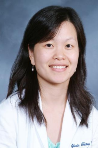
Gloria Chiang, M.D., is an associate professor with the Weill Cornell Medicine Department of Radiology Brain Health Imaging Institute (BHII), and with NewYork-Presbyterian Hospital (NYP). She is a board-certified radiologist with subspecialty certification in neuroradiology. Her research program focuses on combining quantitative magnetic resonance and positron emission tomography (MR) and (PET) imaging techniques with plasma and cerebrospinal fluid (CSF) biomarkers to elucidate the pathophysiology of neurodegenerative diseases.
- Postdoctoral Associate in Radiology
Mohammad Khalafi, M.D., a graduate of Tabriz University of Medical Sciences, reviews medical data and contributes to ongoing research projects at the Brain Health Imaging Institute. Before joining Weill Cornell Medicine, Dr. Khalafi researched neuroradiology and neurodegenerative diseases.
Lidia Glodzik Laboratory
- Assistant Professor in Radiology
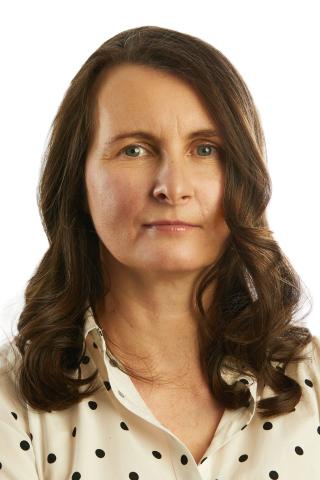
Dr. Lidia Glodzik is the head of the VAscular RIsk in Alzheimer’s disease (VARIA) lab. She completed her residency training in neurology at the University Hospital in Krakow, Poland, where she worked in a stroke unit. She holds a Ph.D. in medical sciences from the Jagiellonian University in Krakow. Her Ph.D. thesis dealt with metabolic changes in post-stroke mood disorders measured with magnetic resonance spectroscopy. She subsequently trained and worked in the New York University Center for Brain Health with Dr. Mony de Leon, a pioneer in Alzheimer’s disease (AD) imaging, on projects related to the prediction of dementia in healthy individuals. Dr. Glodzik employs magnetic resonance imaging (MRI) and positron emission tomography (PET) imaging techniques as well as biofluids to investigate early indicators of cognitive decline.
- Postdoctoral Associate in Radiology
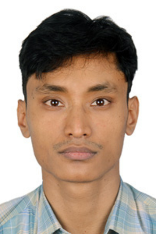
Surendra Maharjan, Ph.D., obtained a doctorate from the Graduate School of Human Health Sciences, Tokyo Metropolitan University, under the Tokyo Human Resources City Diplomacy Scholarship Program. Maharjan received Magnetic Resonance Elastography training in Japan and joined the Indiana University School of Medicine for postdoctoral training. Since then, he has been investigating microstructural and connectivity changes in Alzheimer’s disease mouse models using high-resolution diffusion MRI, honing his expertise in MRI physics, neuroimaging analysis, and applying advanced computational methods in understanding neurological disorders.
At Weill Cornell Medicine, Maharjan processes MR images, including, but not limited to, structural, perfusion, TOF-angio, SWI, and QSM, to extract brain vasculature and tissue property imaging features. He also assists in qualitatively analyzing multimodal images (PET, MRI), proposing experimental designs, and writing manuscripts. Maharjan's research investigates vascular risk factors on brain structure, cerebral blood flow, brain clearance, and the anatomy of cerebral vessels. He's also involved in quantitatively assessing ischemic and neurodegenerative changes in brain aging.
- Assistant Research Coordinator in Radiology
Christopher Mardy received his B.S in biology from the State University of New York, Albany, and later went on to receive his MBS. in biomedical science from Rutgers University, New Brunswick. Christopher is currently pursuing a degree in nursing, with the hope of furthering his research experience with the aging population.
Amy Kuceyeski Laboratory | Computational Connectomics (CoCo) Laboratory
- Professor of Mathematics in Radiology
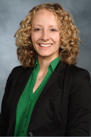
For more than a decade, Amy Kuceyeski has been interested in understanding how the human brain works to better diagnose, prognose and treat neurological disease and injury. Quantitative approaches, including machine learning, applied to data from rapidly evolving neuroimaging techniques, have the potential to enable groundbreaking discoveries about how the brain works. Amy is particularly interested in non-invasive brain stimulation and pharmacological interventions, like psychedelics, that may modulate brain activity and promote recovery from disease or injury. Amy is also the founder and co-director of the cross-campus working group Machine Learning in Medicine, which aims to bring together researchers in Cornell-Ithaca/Cornell-Tech and clinicians/researchers at Weill Cornell Medicine to address medicine's toughest problems. See the group's website.
- Research Associate in Radiology

- Staff Associate in Radiology
Keith Jamison is a staff associate in the CoCo Lab. He has a B.S. in computer science and an M.S. in Biomedical Engineering from Cornell University. Through his education and training, he has developed the broad range of skills and expertise necessary to discern scientifically and clinically relevant patterns from large neuroimaging datasets. While working for the Human Connectome Project (HCP) at the University of Minnesota, he helped implement and adapt preprocessing and analysis pipelines for a large number of anatomical, functional and diffusion magnetic resonance imaging (MRI) scans. He also helped design and test new scanning protocols and modalities for some of the HCP-related studies whose data they now propose to analyze. Since joining the CoCo lab in 2017, he has built upon this expertise in neuroimaging acquisition and processing to help develop modeling approaches using this neuroimaging data to better understand the functional and structural connectivity relationship, and how connectivity relates to both healthy brain function and neurological damage or disease.

- Instructor of Mathematics in Radiology
Ceren Tozlu is a post-doctoral associate in the Weill Cornell Medicine (WCM) Department of Radiology, and in the Cornell University Department of Statistics and Data Science and Computational Biology. She received her M.S. and Ph.D. from the Biostatistics, Biomathematics, Bioinformatics and Health department (3B-H) of Université Claude Bernard Lyon 1 in 2014 and 2018, respectively. She gave lectures to the M.S. students of Cancer, Neuroscience, Biostatistics and Public Health at Université Claude Bernard Lyon for one year and to the medical students at École Santé des Armées (Army Health School of France) for four years. Her M.S. research project focused on application of various machine learning methods to voxel-based conventional human imaging data to predict infarction risk of 3D brain tissue in acute stroke patients. Her Ph.D. thesis focused on modeling disease evolution of stroke and multiple sclerosis (MS) patients based on cross-sectional and longitudinal clinical and imaging data plotted over five years. Her post-doctoral research focuses on (1) modeling of evolution in neurological diseases, particularly in patients with stroke and MS, using statistical and machine learning methods based on demographic, clinical, regional and pair-wise functional and structural connectivity measurements, and (2) identification of the best biomarkers of disease evolution including the particular structural and functional connections contributing to differences in patients with different neurological diseases. Her post-doctoral study aims to develop a novel and personalized model to be used by clinicians to predict individual disease evolution via an application or software, to lead to personalized treatment. She was recently awarded a postdoctoral fellowship grant from the National Multiple Sclerosis Society.

- Ph.D. Student in Electrical and Computer Engineering
Zijin is a Ph.D. student in the Cornell University Department of Electrical and Computer Engineering. She received her bachelor’s degree in electrical engineering from Zhejiang University, China. Her research focuses on the intersection of machine learning and neuroscience. She is particularly interested in applying innovative machine learning algorithms to brain connectivity network analysis. Her project involves developing a noninvasive, spatially unconstrained and personalized method for neuromodulation, which involves creating deep neural networks for stimuli and brain activation patterns mapping. She hopes that manipulating brain connectivity networks will help alleviate symptoms or boost recovery after neurologic injury.

- Ph.D. Candidate in Physiology, Biophysics, & Systems Biology
Lisa is a third-year student in the Weill Cornell Medicine Physiology, Biophysics and Systems Biology Graduate Program, and a recipient of a National Science Foundation Graduate Research Fellowship in bioengineering. After three years of studying economics in construction in her hometown in Russia, she immigrated to the U.S., and graduated with a B.A. in neurobiology from Hunter College in 2019. At Hunter, Lisa also conducted research in the Goldfarb lab, where she tricked brain cancer cells into expressing mutant sodium channels, and then poked them with electrodes to learn how the mutations affected voltage-dependent fast inactivation, a key property of the channel altered in disease phenotypes. Lisa’s strongest aspiration is to contribute to the development of brain-computer interfaces that improve quality of life for people with impaired physical and cognitive function (and maybe even augment natural brain capabilities of healthy people).
Yi Li Laboratory

- Associate Professor in Radiology
Dr. Yi Li, co-director of imaging research for the Brain Health Imaging Institute (BHII), is an associate professor of radiology in the Weill Cornell Medicine (WCM) Department of Radiology. Dr. Li, who trained as a radiologist at Shandong Medical University, earned his doctorate in nuclear medicine from the Shandong University Graduate School. In 2007, Dr. Li became co-director of the New York University (NYU) Center for Brain Health Neuroimaging Laboratory, specializing in structural and positron emission tomography (PET)-related imaging research in aging and dementia. In 2019, Dr. Li’s research team joined WCM to help found the BHII.
- Postdoctoral Associate in Radiology
- Instructor of Biomedical Engineering in Radiology
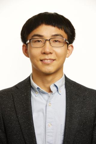
Dr. Liangdong Zhou earned his doctorate in computational science and engineering from Yonsei University. During his Ph.D. program, Dr. Zhou was trained in mathematical and computational modeling of inverse problems in medical imaging, including electrical impedance tomography (EIT), magnetic resonance elastography (MRE), and quantitative susceptibility mapping (QSM). He then joined Dr. Yi Wang's magnetic resonance imaging (MRI) lab—Wanglab—for postdoctoral training in biophysical modeling and magnetic resonance (MR) data acquisition. At Wanglab, Dr. Zhou developed Quantitative Transport Mapping (QTM), a novel perfusion quantification method. By using both MRI and positron emission tomography (PET) imaging, Dr. Liangdong, under the supervision of Dr. Yi Li, is now working on the quantitative analysis of cerebrospinal fluid clearance in neurodegenerative disease.
Laura Beth McIntire Laboratory
- Assistant Professor of Pharmacology in Radiology
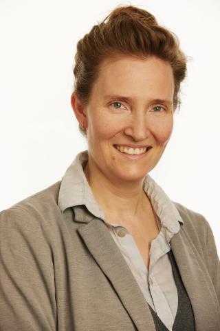
Dr. Laura Beth McIntire, Ph.D., is director of the Lipidomics and Biomarker Lab at the Brain Health Imaging Institute (BHII), and an assistant professor of pharmacology, in the department of radiology. Her work focuses on the contribution of lipid dyshomeostasis to Alzheimer’s disease (AD) with a focus on phosphoinositide metabolism and acyl chain remodeling. Dr. McIntire is an expert in cell biology, cellular imaging and animal models of AD with expertise in animal behavior and the generation of novel mouse strains. She has expertise in lipidomics, imaging mass spectrometry, and system-wide analyses of the lipid interactome which have led to novel insights into lipid deficits in AD progression.
- Graduate Student in Radiology
- Postdoctoral Associate in Radiology
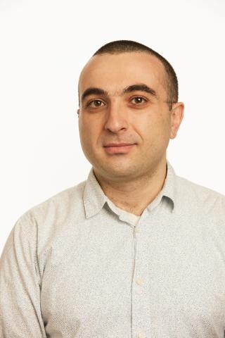
Dr. Artur Lazarian, Ph.D., has extensive experience with mass spectrometry, molecular biology and biochemistry. His recent work has focused on desorption electrospray ionization (DESI) imaging mass spectrometry using the Waters Synapt G2-Si. Additionally, during his previous post-doc position at the Max-Planck Institute for Polymer Research, he extensively used the Waters Synapt G2-Si. In the past two years, he has expanded his expertise to include molecular modeling using Schrodinger software.
Silky Singh Pahlajani Laboratory

- Assistant Professor of Neuropsychiatry Research in Radiology
Dr. Silky Pahlajani is an assistant professor of behavioral neurology in the department of radiology, and an attending physician at NewYork-Presbyterian Hospital. Her primary focus is neurodegenerative disorders, specifically Alzheimer’s disease (AD), and autoimmune antibody-mediated encephalitis, including N-methyl-D-aspartate (NMDA) receptor, leucine-rich glioma-inactivated/contactin-associated protein-2 (LGI-I/CASPR2) antibody, and glutamic acid decarboxylase 65 (GAD-65) antibody, etc. In 2015, Dr. Pahlajani completed her residency training at New York Medical College, where she also served as chief resident. She subsequently completed two fellowships: behavioral neurology/neuropsychiatry at the University of Illinois-Chicago, and central nervous system (CNS) autoimmune disorders at Lenox Hill Hospital, prior to joining the Weill Cornell Memory Disorders Program in September 2017.
Ray Razlighi Laboratory | Quantitative Neuroimaging Laboratory
- Associate Professor of Neuroscience in Radiology
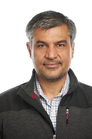
In 2009, Dr. Qolamreza "Ray" Razlighi earned his doctorate in electrical engineering and image processing from the University of Texas. During this time, Dr. Razlighi introduced a new causal Markov random field (MRF) model—Quadrilateral MRF (QMRF)—which has dramatically influenced medical and commercial image analysis, resulting in 12 publications and one patent to date. Dr. Razlighi’s advanced knowledge of neuroimaging and neuroscience started with one year of postdoctoral training at the Brain Imaging Laboratory of the Molecular Imaging and Neuropathology Division, New York State Psychiatric Institute, Columbia University. His education continued with two years of postdoctoral training in the Cognitive Neuroscience Division, Department of Neurology, Columbia University.
Dr. Razlighi has been involved in many neuroimaging projects focused on implementing mathematical models and methods for magnetic resonance imaging (MRI) and functional magnetic resonance imaging (fMRI) data analysis. These include the development of a method for extracting brain features related to the cortical folding pattern; the development of a new non-stationary maximum a posteriori (MAP) QMRF (MAP-QMRF) classifier for brain image segmentation; analysis of fMRI data in a subject’s native space (thereby circumventing the problematic spatial-smoothing step, particularly in studies comparing young/old); and the investigation of inter-hemispheric averaging in resting-state fMRI data analysis. During his postdoctoral training, Dr. Razlighi participated in numerous neuroscience and neuroimaging courses.
- Instructor of Electrical Engineering in Radiology
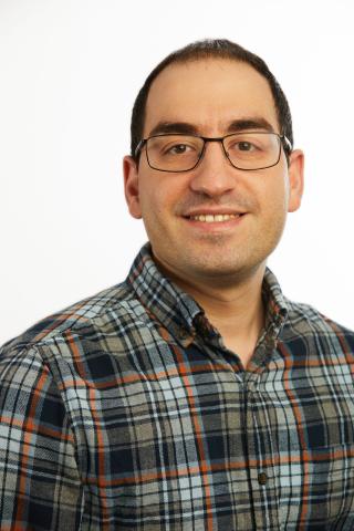
Hani Hojjati, Ph.D., is a postdoctoral associate in the Weill Cornell Medicine Department of Radiology. In 2011, Dr. Hojjati earned his bachelor’s degree in electrical engineering from the Mazandaran University of Iran; from 2011 to 2013, he earned his master's degree in electrical engineering from the Babol Noshirvani University of Technology (NIT); and from 2014 to 2018, he continued his education at NIT, earning a doctorate in electrical engineering.
At NIT, Dr. Hojjati’s thesis focused on predicting Alzheimer's disease (AD) using multimodal neuroimaging methods and machine learning approaches. By employing neuroimaging and multimodal forecasting techniques, he determined accurate predictors for identifying mild cognitive impairment (MCI) versus AD. Using resting-state functional and structural magnetic resonance imaging (MRI) as tools, he enhanced the accuracy of predicting AD.
In 2019, Dr. Hojjati started one year of postdoctoral neuroimaging training at the University of Tennessee Health Science Center Department of Pediatrics. In 2020, he entered a second postdoctoral training program at the Weill Cornell Medicine Department of Radiology. His current research involves using multimodal neuroimaging methods, including structural MRI, and amyloid/tau positron emission tomography (PET), to understand the association between neurodegeneration and amyloid/tau pathologies in the brains of both healthy control and MCI subjects. An award-winning researcher, Dr. Hojjati has published more than 12 journal articles and book chapters and presented at 19 conferences.

- Assistant Research Coordinator in Radiology
A graduate of Daemen College, Sindy Ozoria modeled her individualized studies degree after the history and philosophy of science program at the University of California, Los Angeles. Ozoria, an assistant research coordinator, is interested in applying translational science to vulnerable populations using computational, cognitive, and theoretical neuroscience tools.
Sudhin Shah Laboratory
- Assistant Professor of Neuroscience in Radiology
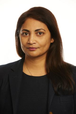
Sudhin Shah, Ph.D., is an assistant professor of neuroscience in the department of radiology within the Brain Health and Imaging Institute at Weill Cornell Medicine (WCM). She received her B.Sc. in electrical engineering from Drexel University followed by a M.Sc. in biomedical engineering from Columbia University. She received her Ph.D. in systems neuroscience from the Weill Cornell Graduate School of Medical Sciences at Cornell University. She completed her postdoctoral training at WCM as well. Dr. Shah is also the Scientific Director of cognitive recovery research at Blythedale Children’s Hospital. She is an affiliate faculty member with the Consortium for the Advanced Study of Brain Injury at WCM. She has served on several National Institutes of Health scientific review panels, and as an invited reviewer for several journals. She has also served as a research mentor for more than 10 medical and graduate students.

- Postdoctoral Associate in Radiology
Ludvik Alkhoury is a postdoctoral associate of radiology at Weill Cornell Medicine. He received a B.S. in electrical engineering from the University of Balamand (UOB), Lebanon. He then joined the Data Fusion Lab (directed by Professor Moshe Kam) at the New Jersey Institute of Technology (NJIT) in 2018. In 2023, he received a Ph.D. in electrical engineering from the Helen and John C. Hartmann Department of Electrical and Computer Engineering at NJIT. His doctoral research focused on wearable health monitoring sensors and methods to extract non-invasive high quality vital signs (such as heart rate and oxygen saturation) during extensive human motion. He is currently working in Dr. Sudhin Shah’s laboratory to study the brain's response to language in children with brain injuries.
- Postdoctoral Associate in Radiology
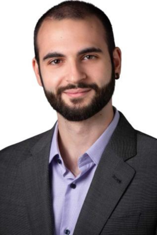
Giacomo Scanavini is a postdoctoral associate in radiology at Weill Cornell Medicine. He holds a Ph.D. in physics from Yale University with a focus on high-energy physics. During his academic journey, he conducted research on neutrino properties, utilizing Liquid Argon Time Projection Chambers (LArTPCs) at Fermi National Laboratory (Fermilab). Currently, Giacomo works in Dr. Shah's laboratory, where he is investigating disorders of consciousness among individuals ranging from young children to adolescents with the goal of developing brain-computer interfaces (BCIs) to enhance essential daily tasks, such as communication.

- Research Assistant
Isabelle Martin is a research assistant at Weill Cornell Medicine. She received a B.A. in Neuroscience and Behavior from Wesleyan University in 2023. She is currently working in Dr. Sudhin Shah’s laboratory and is interested in studying brain injury and its effects on behavior and cognitive abilities.


