The PET Research Laboratory comprises the laboratories of Nikolaos A. Karakatsanis, Ph.D., Associate Professor of Biomedical Engineering in Radiology, and Sadek Nehmeh, Ph.D., Professor of Medical Physics in Radiology.
The research of Nikolaos Karakatsanis, Ph.D., focuses on quantitative whole-body dynamic PET to advance the clinical adoption of multi-parametric PET imaging, enhancing diagnostic accuracy and therapy response assessments in oncology, cardiology, and neurology.
Dr. Karakatsanis has developed dynamic PET-driven quantitative methods to improve attenuation correction and dual-radiotracer decoupling in simultaneous PET/MR imaging, primarily for cardiovascular disease evaluations. He is developing cost-effective long axial field-of-view (LAFOV) sparse PET detection systems to expand clinical access to total-body dynamic PET imaging.
He has co-authored over 50 peer-reviewed original articles and 80 conference records in national and international scientific meetings. A member of the Society of Nuclear Medicine and Molecular Imaging, he served as an intern from 2017 to 2019 — then later as a secretary officer from 2019 to 2021 — in the Physics, Data Sciences, and Instrumentation Council. During his internship, he founded NMMItools.org, a web resource page compiling validated commercial and open-source software tools for nuclear medicine and molecular imaging research.
Dr. Karakatsanis is a senior member of the Institute of Electrical and Electronics Engineers (IEEE), where he served as member-at-large from 2017 to 2020, and has been the secretary officer of the IEEE Nuclear Medical Imaging Sciences Council since 2021. He is certified by the American Board of Science in Nuclear Medicine in nuclear medicine physics and instrumentation. Additionally, he is an associate editor for Frontiers of Medicine and Clinical Imaging and serves as a guest editor for Medical Physics.
The Karakatsanis Lab is designing clinically adoptable dynamic whole-body human PET scan protocols tailored for short AFOV human PET scanners, which are widely available today. These protocols aim to extend multi-parametric imaging benefits beyond static SUV PET imaging to oncology, cardiology, and multi-organ — for example, brain-heart axis — studies.
Having pioneered the first dynamic whole-body 18 FDG PET/CT human scan protocols, the lab now collaborates with nuclear medicine and radiology physicians to expand and customize these protocols for other key radiotracers, including 68Ga-PSMA and 68Ga-DOTATATE, to enhance diagnosis and treatment response assessment for metastatic castrate-resistant prostate cancer, neuroendocrine tumors, and cardiovascular diseases.
The lab is investigating novel dual-radiotracer PET/MR acquisition protocols, leveraging 18F-NaF as a uniquely dynamic agent for bone segmentation and enhanced tissue attenuation mapping. This approach improves attenuation correction in PET/MR studies, compensating for the absence of CT-based attenuation correction, the PET imaging gold standard.
To advance multi-parametric whole-body PET imaging, even with limited AFOV scanners, the lab has developed direct 4D PET image reconstruction algorithms optimized for multi-bed and continuous bed-motion acquisitions. These algorithms integrate graphical analysis methods, allowing for high-precision imaging of multiple clinically relevant macro-kinetic parameters, despite the high noise levels in dynamic whole-body PET data.
A key advantage of these methods is their ability to significantly shorten the total scan period required to capture the macro-dynamics of the administered radiotracer. The lab aims to align scan periods post-injection (PI) with those used in standard-of-care static PET scans — for example, 60–80 min PI for whole-body 18F-FDG PET exams — without compromising kinetic macro-parameters accuracy and precision.
To further streamline dynamic PET imaging, the lab is building population-based input function models for various radiotracers. By analyzing arterial blood plasma concentration data from past dynamic PET exams, these models allow the lab to prospectively acquire only a small portion of the input function at later time points, inferring missing early-phase data from the population model. This innovation brings dynamic PET closer to routine clinical adoption.
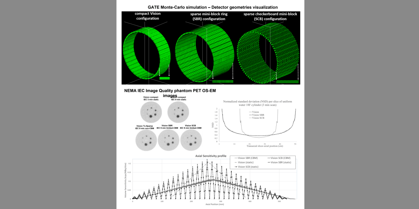
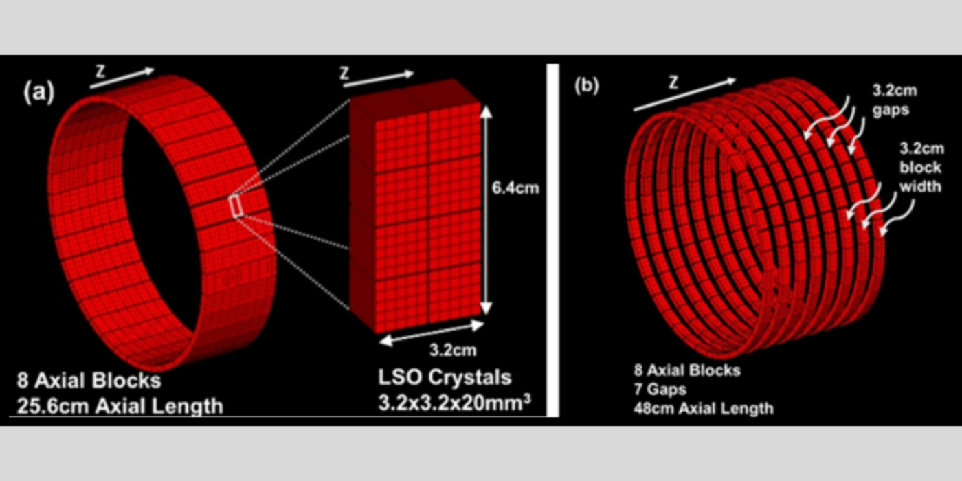
The Karakatsanis Lab is pioneering ML-aided image reconstruction for cost-effective, sparse PET detector geometries with double the AFOV of conventional compact PET scanners. To ensure high-quality image reconstruction despite gaps in detector coverage, the lab developed a customized component-based normalization method to account for sparse sampling effects.
Additionally, the lab introduced a limited-CBM PET acquisition mode to smooth axial sensitivity variations between detector blocks and large gaps. In collaboration with other WCM AI experts, the lab explores U-net deep convolutional neural networks to recover image quality losses and mitigate noise in sparse PET data.
These advances could enable the development of adaptable, cost-effective clinical PET scanners with adjustable AFOVs. Future PET scanners may dynamically configure themselves for high-sensitivity scans over shorter AFOVs or moderate-sensitivity scans over longer AFOVs, optimizing imaging performance based on specific research and clinical needs.
Sadek Nehmeh, Ph.D., has extensive experience in PET physics, clinical PET imaging, and Monte Carlo simulations. His PET physics research focuses on Monte Carlo simulations for novel LAFOV PET systems, application-specific PET systems for noninvasive arterial input function imaging, and Compton scatter PET.
Clinically, Dr. Nehmeh’s research spans hypoxia imaging, simultaneous multi-tracer PET imaging in glioblastoma, PET imaging of multiple sclerosis, cardiac PET imaging, cardiac toxicity in cancer patients, and Alzheimer’s disease. He’s also focused on treatment response assessment in both cancer and neurology.
The author of some 70 publications and book chapters, Dr. Nehmeh has received multiple National Institutes of Health (NIH) grants, industry-sponsored awards, and institutional funding. He also serves as a co-investigator on numerous NIH awards.
Dr. Nehmeh, an active member of the Institute of Electrical and Electronics Engineers, Society of Nuclear Medicine and Molecular Imaging, and American Association of Physicists in Medicine, frequently presents his work at international and national conferences.
He is the director of a WCM medical imaging nuclear physics course, is certified by the American Board of Radiology (ABR), and holds a medical nuclear physics license from the state of New York.
Mony de Leon, Ed.D.
Howard Fine, M.D.
Edward K. Fung, Ph.D.
Susan Gauthier, D.O., M.P.H.
Jana Ivanidze, M.D., Ph.D.
Nikolaos A. Karakatsanis, Ph.D.
James Kelly, Ph.D.
Gene Kim, Ph.D.
Rajiv S. Magge, M.D.
Lisa Mosconi, Ph.D.
 (a) and corresponding hypoxic GTV (GTVh) by 18 fluorodeoxyglucose (FDG)-positron emission tomography (PET) (b-red) and 18F misonidasole (MISO)-PET (b-blue) of a locoregionally advanced supraglottic carcinoma. Corresponding 18FMISO-PET–guided intensity-modulated radiotherapy plan (c). Dose to hypoxic target volume escalated from 70Gy to 84Gy.")
