The Gene Kim Lab is focused on understanding the tumor microenvironment through magnetic resonance imaging (MRI), and developing efficient non-invasive imaging tools for reliable prediction and assessment of treatment response in cancer. Treating aggressive cancer remains challenging. It is often unclear whether the lack of an effective response is due to a failure to deliver therapeutics, or an ineffectiveness of therapeutics to kill cancer cells. It is therefore necessary to assess both blood flow and cell death to elucidate the underlying mechanism, and to optimize the treatments. The overarching goal of our research is to understand the link between MRI measures and tumor microstructural and functional properties. Understanding these properties can be beneficial to the development of new diagnostic and therapeutic approaches.
To unravel the complexity of cancer, the lab has been working on several MRI studies with a focus on (i) tumor vascularity, (ii) cellular properties, and (iii) the association of adipose tissue with cancer development and treatment.
Associated Lab Members

Dr. Gene Kim is a professor of biomedical engineering in radiology at Weill Cornell Medicine. He received his Ph.D. in biomedical engineering from the University of Southern California and completed his postdoctoral fellowship in cancer imaging at the University of Pennsylvania. His research focuses on the development of quantitative dynamic contrast-enhanced and diffusion magnetic resonance imaging methods for early detection of cancer and assessment of treatment response, particularly in breast cancer and head and neck cancer. Dr. Kim’s laboratory has been funded by grants from the National Institutes of Health.

Miran Jang holds a bachelor’s and master’s in bio and brain engineering from KAIST (Korea Advanced Institute of Science and Technology). Throughout her master's program, she developed an innovative microfluidics device for single-algal-cell cultivation. After completing her master’s degree, Miran joined the analytical team at Celltrion R&D Center, a leading biologics company. Her research expertise revolves around developing physical and chemical analysis assays for antibody biologics. She has experience developing antibody drugs, from early development to final approval. She has managed and led major BLA (Biologics License Applications) studies for biosimilars and a new drug.

Dr. Seung Koo Lee is an assistant professor of cell biology research in the Weill Cornell Medicine (WCM) Department of Radiology. His research focuses on the development of, (1) innovative multi-functional molecular imaging probes for various disease states including cancers and, (2) drug delivery systems for diagnostic, therapeutic and clinical applications. He earned his Ph.D. in immunology from Seoul National University, where he studied the signaling mechanisms of vitamin C that control tumor proliferation, and apoptosis in colon cancer and melanoma. He then completed postdoctoral training at the Houston Methodist Research Institute under the guidance of Dr. Ching Tung, where he invented a layer-by-layer nanoplatform (LbLN) that delivers a short interfering RNA (siRNA) and imaging probe to various cancers.

Sawwal Qayyum received his bachelor’s degree in biology from Ramapo College of New Jersey. During his undergraduate training, he spent his seminar year looking at embryogenesis-lethal genes in C. elegans using RNA interference (RNAi) and clustered regularly interspaced short palindromic repeats (CRISPR)/Cas9 to visualize protein localization via green fluorescent (GFP) recombinant vectors. He previously worked at the New York University Langone Medical Center as a senior animal care technician, attaining there his laboratory animal technologist certification. He spent two years interning at the Preclinical Imaging Core headed by Dr. Wadghiri. There he became familiar with different modalities of optical imaging, magnetic resonance imaging (MRI), and tumor pH probe design. In the Gene Kim lab, Sawwal is studying the effect of metronomic chemotherapy on the tumor vasculature and angiogenesis of orthotopic triple-negative breast cancer (TNBC) and glioma mouse models using dynamic contrast-enhanced (DCE)-MRI.

Myung-Shin (Cindy) Han holds a master's in Molecular Biology from Sookmyung Women's University in Seoul, Korea. After finishing her master’s, Cindy worked as a research manager in orthopedic surgery at Seoul National University Hospital, researching tumor immunity and stem-cell therapy with bone grafts. At the Methodist Hospital Research Institute in Houston, Texas, as a research assistant, she researched molecular optical imaging with tumor cell lines in animals. Since 2013, she's worked at Weill Cornell Medicine. For over 20 years, Han has had myriad experiences in animal experiments, and her expertise extends to innovative nanomedicine for tumor imaging and therapy. She's also working with sprayable fluorogenic dyes for image-guided ovarian cancer surgery.

Eddy Solomon’s research combines diverse aspects of magnetic resonance imaging (MRI), including pulse sequence programming, advanced image reconstruction, and pre-clinical and clinical patient studies. He received his Ph.D. from the department of chemical physics at the Weizmann Institute of Science, where he developed novel MR methods, based on spatiotemporal encoding (SPEN) principles. He is now a Weill Cornell Medicine faculty instructor of chemical physics in the radiology department. He works on head and neck cancer using diffusion and dynamic contrast-enhanced (DCE) approaches. His aim is to advance body magnetic resonance imaging (MRI) methods in humans, to improve both basic knowledge and patient care.
Imaging tumor vascular properties
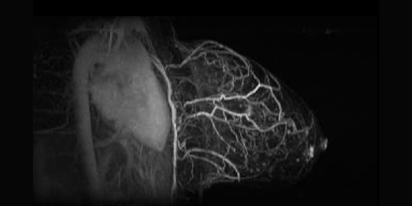
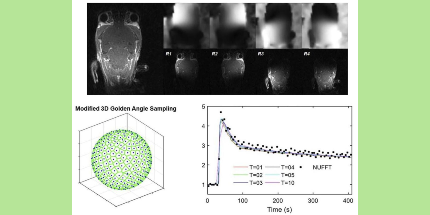
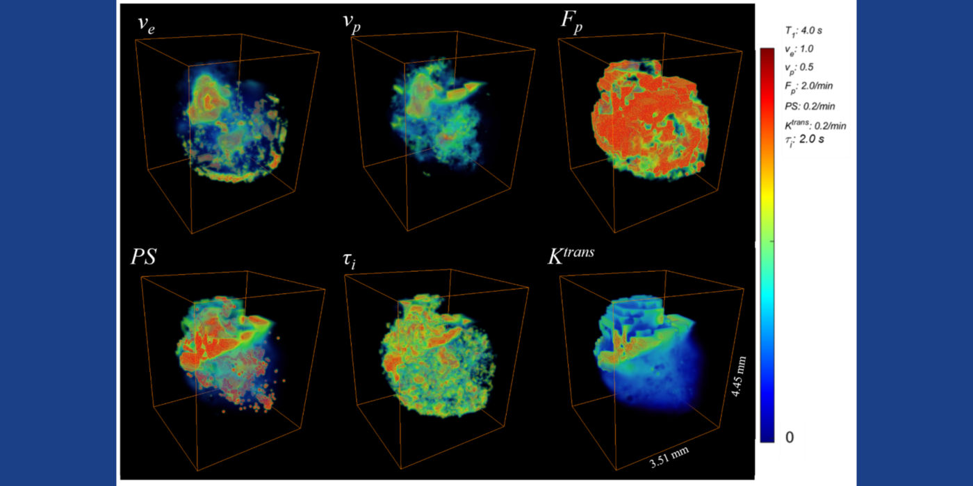
Chaotic vascular growth is a hallmark of malignant tumors and an important target for cancer treatment. The primary method that we have been using for the study of tumor vascularity is dynamic contrast enhanced (DCE) MRI (Kim et al., 2007 and 2010). In order to further understand the contrast dynamics inside the tumor with a high precision, the lab has been actively developing fast MRI methods that provide a higher temporal and spatial resolution, including the 3D ultra-short echo time (UTE) golden-angle radial sparse and parallel (GRASP) MRI method (Feng et al., 2014; Zhang et al., 2019).
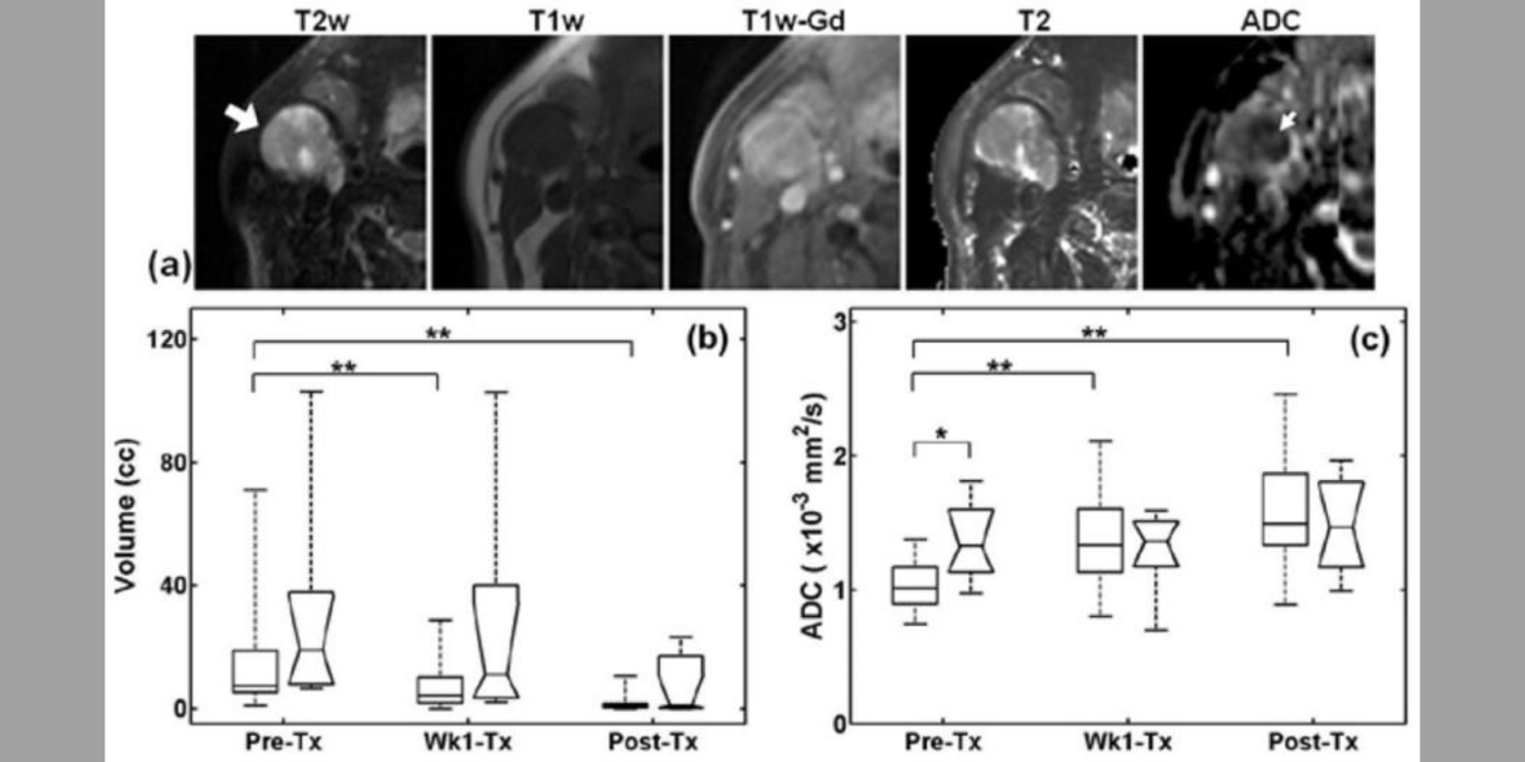
Among many contrast kinetic parameters that can be measured using DCE-MRI, the Kim lab is particularly interested in the cellular-interstitial water exchange rate. The lab's recent head and neck cancer study found patients with slower transcytolemmal water exchange rates at pre-treatment have significantly prolonged overall survival at five years and beyond (Chawla et al., 2018). The lab showed that the effect of water exchange can be actively encoded in the dynamic data in order to improve the precision of water exchange rate measurement (Zhang and Kim, 2019). In addition, the lab introduced a single comprehensive imaging method, namely active contrast encoding (ACE)-MRI (Zhang et al., 2017), that provides pre-contrast T1 and actual flip angle from the dynamic data and eliminates the need to measure them separately. The goal: bring quantitative DCE-MRI to routine clinical imaging exams for accurate assessment of cancer treatment response, and also to extend its use to other microvascular diseases.
Imaging tumor cellular properties
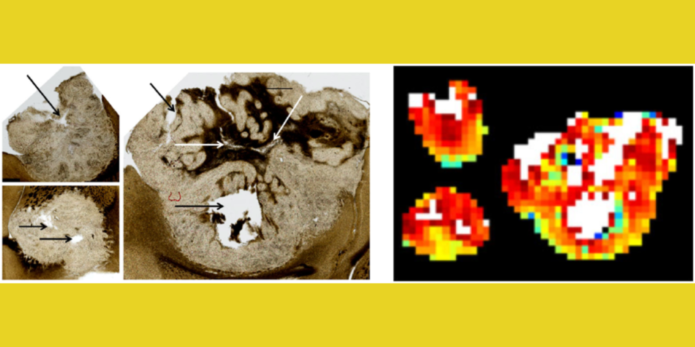
Another useful approach for the assessment of tumor treatment response is to measure cellular microstructural properties using diffusion MRI (dMRI) (Kim et al., 2009). Conventional dMRI measures such as apparent diffusion coefficient, however, remain non-specific biomarkers. The dMRI technique allows one to represent different biophysical properties of tissue depending on the diffusion weighting strength (q) and diffusion time (t) used for the measurement. The feasibility of measuring cell size using the t-dependency of dMRI was reported earlier with reference to different muscle groups (Kim et al., 2005). Inspired by these results, the lab has focused on investigating quantitative ways to utilize both the q- and t-dependency of dMRI data for assessment of cell viability by measuring cell size, extracellular volume fraction, and cellular compartmental diffusivities. This led the lab to propose the POMACE (pulsed and oscillating gradient MRI for assessment of cell size and extracellular space) framework (Reynaud et al., 2016) as a non-invasive imaging method to measure cellular microstructural properties.
Moreover, the lab has demonstrated that t-dependency of diffusional kurtosis can also be used to measure the cellular-interstitial water exchange rate (Zhang et al., 2021). This innovative approach does not require the use of an exogenous contrast agent to measure the water exchange rate and can provide a non-contrast imaging marker for cellular metabolism. The lab's investigation of quantitative dMRI measures in cancer, including the cellular-interstitial water exchange rate in head and neck cancer patients undergoing radiation therapy, is conducted in collaboration with the National Institutes of Health/National Cancer Institute (NIH/NCI) Quantitative Imaging Network.
Association of adipose tissue with cancer development
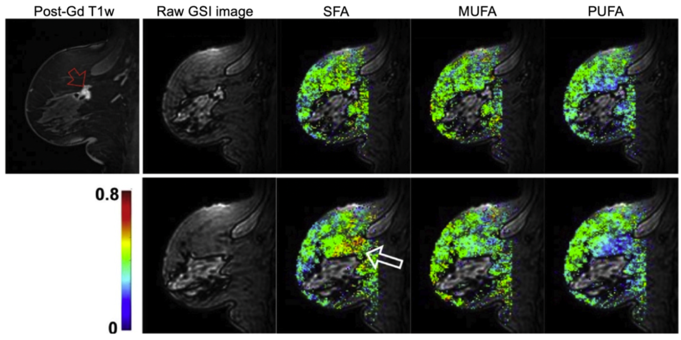
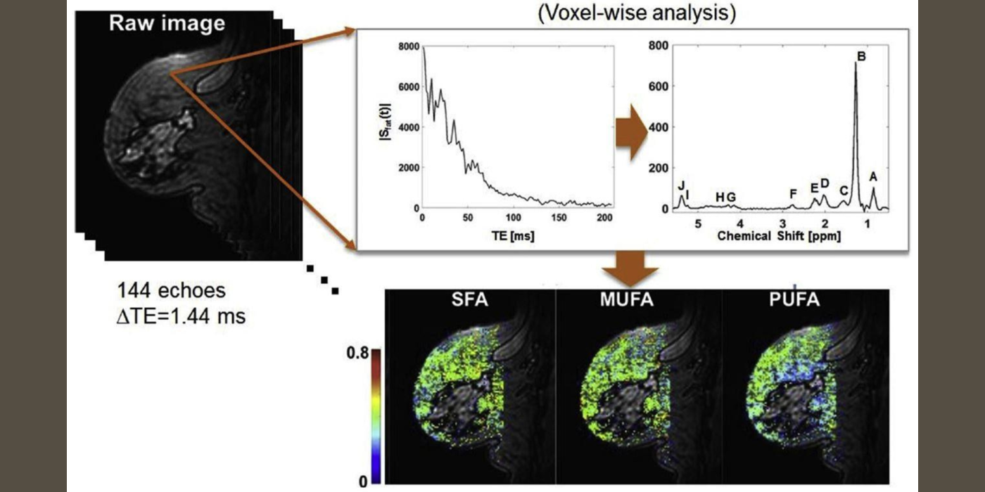
It has been increasingly recognized that adipose tissue may not be an “innocent bystander” in cancer development and progression. The lab has been developing fast MR spectroscopic imaging methods to measure fatty acid compositions in breast adipose tissue and investigating how these fatty acids are associated with breast cancer (Freed et al., 2016). After menopause, adipose tissue becomes the principal site of estrogen biosynthesis, and there is ample evidence demonstrating possible links between tissue hormone levels and fatty acid composition. While these hormonal effects are recognized as significant confounding factors in breast MRI, they may also provide a non-invasive means to study the effect of hormones on normal breast tissue and its association with breast cancer development and progression as demonstrated in our previous studies (Amarosa et al., 2013; Clendenen et al., 2013).
The team aims to develop MR spectroscopic imaging methods to measure fatty acid composition in breast adipose tissue and to assess its association with female hormones at the tissue level; the vascular and cellular properties of breast fibroglandular tissue; and cancer development.
Award or Grant: R01CA219964, National Cancer Institute (NCI) (02/01/18 – 01/31/23).
Description: The role of breast fatty acid composition in breast cancer development, in conjunction with breast density and body mass index, has not been fully investigated. The central hypothesis of the Kim lab is that...
Awards or Grants: UG3/UH3CA228699, National Cancer Institute NCI, 5/1/2019 to 4/30/2024
Description: Quantitative diffusion magnetic resonance imaging (dMRI) remains challenging as dMRI data represent different biophysical properties of tissue depending on diffusion weighting strength (q) and diffusion...
Awards or Grants: R01 CA160620, National Cancer Institute (NCI), 2/1/2019-1/31/2024
Description: Assessment of cancer treatment requires an effective non-invasive method of measuring both the vascular and cellular changes induced by therapies. The Kim team’s underlying hypothesis is that a single...