Citigroup Biomedical Imaging Center (CBIC)
 The Biomedical Imaging Core Facility at the Citigroup Biomedical Imaging Center houses hardware, software, and professional support for a range of imaging modalities including magnetic resonance imaging (MRI), magnetic resonance spectroscopy (MRS), positron emission tomography (PET), computed tomography (CT), single photon emission computed tomography (SPECT), multispectral optical imaging, and ultrasound. In addition, a fully supported cyclotron and radiochemistry facility is devoted to the synthesis of radiotracers for a wide range of applications. The entire facility is dedicated to biomedical imaging research and applications development.
Pre-clinical Imaging
The small animal imaging facilities at CBIC consist of an Inveon multi-modality PET/CT/SPECT system, a Bruker Biospec 7.0 Tesla 30 cm bore magnetic resonance imaging spectrometer, a Bruker Xtreme multi-spectral imaging system, two Scano micro CT units, a Perkin Elmer IVIS optical/CT system, a Siemens CTI Focus 220 PET system, and a Visulsonics Vevo 3100 ultrasound system. The scanners are supported by immediately adjacent space and staff for animal preparation including surgical capabilities. In addition, our human subjects MRI hardware has been modified in-house to accommodate small animal imaging, thereby enhancing translational imaging capability.
(1) PET, SPECT, and CT:
The Siemens Inveon preclinical imaging platform provides integrated small animal PET, SPECT and CT imaging and analysis. Inveon can be used as a fully integrated (PET/SPECT/CT) or dockable system with single or multimodality configurations. Networked computers embedded in the gantries coordinate PET, SPECT, and CT data acquisition.
The Siemens microPET Focus 220 preclinical PET scanner is a large-bore PET system designed specifically to accommodate a range of animal sizes, allowing to visualize and quantify disease and the action of novel therapies in living animals. It features a 22 cm bore size, 1.4 mm resolution, easy access to animals, and continuous bed motion. A bed pallet provides effective axial FOV of 50 cm and support for animals up to 25 kg in weight. Whole-body imaging with continuous bed motion enables applications such as biodistribution analysis and dynamic imaging.
The Scanco VivaCT 40 offers a fast, flexible platform for in vivo studies of mice. Both whole body scans and scans from selected regions of interest are supported. A range of beds is included with the scanner allowing for both in vivo and specimen studies. The Scanco mCT 35 complements the VivaCT 40 and is a desktop ultra high resolution specimen system offering a spatial resolution as high as 10 microns. Both units are fully self-shielded, allowing for simultaneous use in their installed location.
(2) MRI and MRS
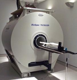 The Bruker Biospec 7.0 Tesla magnetic resonance imaging (MRI) spectrometer is equipped with a 12 cm diameter 450 mT/m actively shielded gradient subsystems with integrated shim capability. The system features several linear and circularly polarized radiofrequency resonators for imaging of small animals and includes double tuned
1H/31P and 1H/13C coils for spectroscopy. In addition, we have designed several high sensitivity radiofrequency resonators for this system in-house, including one that is incorporated into a home-built magnetic resonance microscopy insert. The system supports parallel imaging, and an animal monitoring system is included. Also, this system has been modified to allow for small animal functional MRI and includes a stimulus isolation unit with an arbitrary wave form for somatosensory stimulation experiments, an MRI compatible loudspeaker system for auditory stimulation experiments in songbirds and marmosets, and optogenetic fMRI capabilities.
(3) Multi Spectral Optical/X-ray imaging:
Our new Bruker Xtreme multi-spectral imaging system and Perkin Elmer IVIS optical/CT offers quantitative imaging of luminescent, radioisotopic,and multi-wavelength fluorescent labeled biomolecules in combination with radiographic imaging for small animals. They feature up to 10 micron/pixel resolution.
(4) Ultrasound Imaging:
The Vevo 3100 combines ultra high-frequency imaging, quantification and education in an all-in-one platform designed for small animal studies in a biosafety level-2 compliant environment. The Vevo platform contains a high-frequency array-based ultrasound imaging system that enables in vivo anatomical, functional, physiological and molecular data simultaneously, in real-time and with a resolution down to 30 microns. The system is easy to use, non-invasive and fast, thus providing extremely high throughput.
Human Subjects Imaging
Our translational and human subjects research imaging hardware is located in the same building as most of the small animal imaging systems. Platforms include both Siemens PRISMA and General Electric Discovery MR750 3.0 Tesla MRI/MRS systems, and a Siemens Biograph PET/CT system.
(1) PET/CT
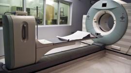 The Siemens Biograph mCT 64-slice PET/CT tomograph is designed for oncological, neurological and cardiac imaging. It consists of a multi-LSO-detector ring system with 3D acquisition and reconstruction, and 81 image planes with a 16.2 cm axial field of view. It has a 78 cm gantry aperture, 70 cm transverse field of view and 16.2 cm axial field of view with fast acquisition and reconstruction of 128 x 128 and 200 x 200 matrices. It has static, whole-body, and list mode acquisition capability. The 500 lb capacity bed allows imaging of nearly all subjects. The 64-slice CT imaging capability consists of a full range of SPIRAL CT clinical applications.
(2) MRI
Our Siemens 3.0 Tesla PRISMA MRI/MRS system is a 60 cm bore top-of-the-line research product featuring 64-channel operation with 102 channel plug-ins. It also features an 80 mT/m 200 T/m/s gradient for rapid imaging applications. The system includes a full multinuclear spectroscopy platform as well as full advanced cardiac capability. A wide range of radiofrequency resonators allows for full body coverage including both 32- and 64-channel head coil, spine array, body phased arrays, and extremity arrays. Investigator built resonators and pulse sequence development platforms are supported. In addition we have modified this system in-house to allow for state-of-the-art functional magnetic resonance imaging acquisition. Functional MRI paradigms are supported with a full complement of hardware and software research tools. Eye tracking capabilities for both human and primates are provided with real time monitoring.
The General Electric 3.0 Tesla Discovery MR750 MRI/MRS System is also a top-of-the-line research product that features an array of high performance accessories for high resoluton anatomical and functional imaging, and includes multinuclear spectroscopy and full advanced cardiac capability. A SensaVue fMRI stimulus delivery system features a battery of validated fMRI tasks that provide proven activition of the motor, visual, auditory, and language regions of the brain. A comprehensive set of specialized radiofrequency resonators has been built in-house for pre-clinical imaging. The small animal resonators greatly aid in translational research since the same pulse sequences can be applied directly to human subjects. Research developed pulse sequences are also supported.
(3) Magnetic Resonance Guided Focused Ultrasound:
Our General Electric Discovery MR750 MRI/MRS system is equipped with an Insightec ExAblate Neuro magnetic resonance guided focused ultrasound (MRgFUS) platform for applications in the brain. The ultrasound tables are dockable to the MRI system, and the system allows for digital control of the amplitude and phase of each transmit channel. The ExAblate Neuro is fully integrated with the MRI system in that both localization images for treatment planning and temperature maps for treatment verification are provided via MRI.
Cyclotron and Radiochemistry
The cyclotron facility consists of an EBCO TR-19 dual beam, negative ion (19 Mev proton and 9 MeV deuteron at <150 µAm) self-shielded cyclotron with 8 different target ports. The cyclotron has the basic targetry for the production of F-18 (>2Ci), C-11 (1.5 Ci) and N-13 (1.2 Ci and NH3). In addition, the external beam-line was designed to support a semi-automated solid target system capable of making I-124 and Zr-89. The cyclotron is located in the same building as the PET cameras and facilitates the use of short -lived isotopes such as O-15 and C-11.
The radiochemistry laboratory facility is equipped with 3 hot cells and 2 mini cells. The lab has 2 automated [11C]Methyiodide and methylation modules ((GE TracerLab FXc pro and GE MicroLab) for the production of [11C]CO2, [11C]Methyliodide and [11C]methyl triflate for the routine synthesis of IND PET radiotracers such as [11C]PIB, [11C]Raclopride, [11C]Arachidonic acid, [11C]Flumazenil and [11C]PK11195. In addition the lab is equipped with automated synthesis modules for the preparation of 68Ga labeled peptides. The Quality Control lab is fully equipped to perform routine quality control testing for final drug product release for patient studies. The equipment includes; GC, HPLC, an ITLC-scanner, an MCA and an automated well counter. In addition, the microbiology lab is set-up to perform, endotoxin and sterility testing, environmental monitoring, growth promotion testing, and media fills. The manufacturing and QC testing labs are cGMP compliant, and are equipped with an aseptic environment (2 Laminar work benches) for the production of PET radiopharmaceuticals according to FDA 21 CFR Part 212 regulations.
Image Processing and Programming Infrastructure Deployed within CBIC
Hardware:
- >20x Windows PCs (Windows XP/Windows 7)
- 3x Linux PC's (Fedora, SUSE, etc.)
- 2x Apple Mac Pro
- 2x GE PACS Workstations
- 2x Siemens Leonardo Workstations
- 2x GE Entegra PET/CT Workstations
- 1x Apple Mac Book
- 1x Buffalo 8Tb Network Attached Storage Terastation Server
Image Processing and Programming Tools:
- 4x MATLAB R2011 Licenses
- 3x IDL 8.1 Licenses
- 2x PMOD 3.3 PET Modeling Software
- PBAS - Base incl. PVIEW, DICOM server, DB, key
- PKIN - General kinetic modeling tool, with all models
- PXMOD - Pixel-wise modeling tool, with all models
- PCARD - Cardiac modeling tool (Extension)
- PFUS - Image registration and fusion tool
- P3D - 3D image rendering tool
- PALZ - Alzheimer discrimination tool
- 1x General Electric MRI Pulse Sequence Compilers
- 1x Siemens MRI IDEA Pulse Sequence Compilers
- 1x BrainVoyagerQX 2.3
- 1x EPrime fMRI license
Imaging Informatics and Infrastructure to Support Clinical Trials / Human Subjects Research
Image Data Evaluation and Analytics Laboratory (IDEAL)
 The Image Data Evaluation and Analytics Laboratory (IDEAL) has clinical, research, and research infrastructure foci. IDEAL provides clinical image post-processing support for the Department of Radiology and its referring physicians. In addition, the lab has its own research mission with active projects in memory disorders (including NPH, AD, MCI, PD); movement disorders (including MS, PD); CT dose reduction (including screening, HRCT, body composition, CT Perfusion, oncologic imaging); enhanced post-processing technology development (including segmentation, registration, dataset fusion, lesion characterization). Finally, IDEAL has a significant mission in providing research infrastructure including the institutional Research PACS (serving WCMC, the CTSC, the Cancer Center, and other entities), providing clinical trials management services for Phase 0 – IV trials, and providing research interpretations (such as RECIST, Cheson, arthritis scores, etc.).
IDEAL has developed clinical tools such as a Brain Health Report Card that includes measures of CSF flow, perfusion, morphometry, brain diffusivity, and quantitative susceptibility mapping. IDEAL has also developed research support tools such as STARS (Study/Subject Tracking And Registration System) and semi-automated measurement tools.
Hardware:
- 8 PCs running Windows XP / Windows 7, 4GB RAM, 3GHz dual-core and quad-core Intel processors
- 7 desktop machines running under Ubuntu-Linux or dual boot Linux/Windows
- 1 Linux server with expandable 1.2 terahertz hard drive, running open source research PACS
- 1 PC dedicated to the TeraRecon™ 3D “thin client” visualization and processing software
- 2 GE Advantage Workstations for advanced 3D imaging and processing
- 2 Laptops running Windows 7
- Apple Mac Pro (8 cores, 16 GB RAM)
- 1 GE AWS with the full suite of analysis tools (version 4.4)
- TeraRecon iNtuition thin client clinical server, with 12 TB storage
- TeraRecon thin client research server, with 17 TB storage
- Brainlab thin client clinical and research server
- Drobo server for backup and longer term storage for research data only. The Drobo is an 8-bay DroboPro-FS with nominally 19 TB of hard drives (five 2TB, three 3TB) and in the current set up with dual-disk redundancy has 13 TB (11.8 TiB) of storage space available. (1 TB = 10^12 = 1000^4 bytes; 1 TiB = 2^40 = (1024)^4 bytes; each disk of redundancy loses one copy of the largest drive 19TB-3TB-3TB=13TB). It has two gigabit ethernet connectors, however we are currently using only a single connection at 100Mbps. It operates as a CIFS / NFS / SSH / SFTP network file server.
- We maintain a mirror computational development, validation, and optimization facility at the Robotics lab on the Cornell University main campus in Ithaca available for the use of CS undergraduates, MEng students, and PhD candidates with access to the WCMC network; this mirror facility includes 3 PCs running under both Windows and Linux.
Image Processing and Programming Tools:
- 8x MATLAB R2011 Licenses (Win 32, Win64, iOS, Linux)
- 3x FreeSurfer Installations (Open Licenses)
- 4x FMRIB Software Library (FSL) for Neuroimaging analysis in Linux
- 6x Statistical Parametric Mapping (SPM) Installations (Open Source MATLAB Toolbox)
- 2x Microsoft Visual Studio Suite for software development in C/C++
- 4x Image ToolKit (ITK) C++ library Installations (Open Source)
- 4x Visualization ToolKit (VTK) Lifetime Licenses
- 3x TrackVis DTI Tractography software suite installations (Open Source)
- Remote access to Vital Vitrea workstation
- Full suite of TeraRecon post-procesing tools
- Full suite of GE AWS v4.4 tools on two IDEAL dedicated workstations (additional workstations throughout enterprise for clinical use)
- OsiriX current version
MR Spectroscopy Lab (Shungu, and Mao)
Hardware:
The laboratory is equipped with a dual-processor (2 x 900 MHz) Sun Microsystems Incorporated (SMI) Blade 1000 workstation, a dual-processor (2 x 360 MHz) SMI Ultra 60 workstation, a uniprocessor SMI Ultra 2 workstation, two uniprocessor SMI Ultra 10 workstations, and a uniprocessor SMI SparcStation 10 workstation. A 2.4 GHz Dell Optiplex 210L running under the LINUX operating system and equipped with several versions of the GE Environment for Programming in C (EPIC) and appropriate compilers is available for pulse sequence development and simulations. All of these workstations are interconnected in a local area network (LAN), in which they exchange and share resources, and are also connected to all the MRI units. Assorted Windows laptops and desktop personal computers, various statistical software packages (e.g., SPSS), word processing (MS Word, Adobe FrameMaker), graphical and imaging processing (MEDx, SPM, AFNI) software, as well as hardcopy devices (laser printers, faxes, copiers, scanners) are also available.
Image Processing Tools:
XsOsNMR, Dr. Shungu’s laboratory has developed a versatile, intuitive and full-featured IDL-based software package for processing, analyzing, and displaying in vivo single-voxel, and multidimensional spectroscopic and structural imaging data. The software is sufficiently automated to be used by individuals with little experience, but it is also equipped with advanced features which allow experienced users to interactively process all forms of 1D, 2D, 3D, and 4D MRS and conventional anatomic MRI data. This software’s spectral analysis tool, which is being updated to permit automated absolute metabolite quantification, is fast, reliable and has shown a great deal of stability in the analysis of thousands of spectra over the past decade by us and by collaborators and colleagues at other institutions (e.g., Johns Hopkins School of Medicine, University of Pennsylvania, Cornell Weill Medical Center, Memorial Sloan-Kettering, Columbia University College of Physicians and Surgeons).
MRI Research Institute (Wang, Spincemaille, Nguyen, Prince, et al)
Hardware:
- 16x Windows PCs (>8GB RAM each)
- 6x Linux PCs (file server/GE psd compilers)
- 2x Apple Mac Pro (8 cores, 16 GB RAM)
- 2x Apple MacBook Pro
- 32 compute node cluster (3.2 GHz P4 Xeon EM64T, 6 GB RAM, Dell PowerEdge 1850)
- 1x Dell R910 (8x8 cores, Intel® Xeon® X7550, 2GHz, 64 GB RAM)
- 1x Dell R910 (4x8 cores, Intel® Xeon® X7550, 2GHz, 64 GB RAM)
Image Processing and Programming Tools:
Software available in this lab includes OsiriX medical image viewer (both commercial and free versions, including several in-house developed plugins), MATLAB, LVMETRIC for automated left ventricle segmentation of cardiac MR images, SPM, and NIFTI. Development tools include GCC and Intel C/C++ compilers, GE epic pulse sequence compiler, ATLAS, MKL, DCMTK, and in-house developed tools and libraries for GE MR raw data processing, real-time MR navigator, MR image reconstruction, and magnetic susceptibility mapping.
Dalio Institute of Cardiovascular Imaging (Mosadegh, Dunham)
Fabrication Equipment:
- 1 Stratasys J826 Multi-material 3D printer
- 1 DLP-based 3D printer
- 1 Extrusion-based 3D printer for liquid materials (custom-built)
- 1 Laser cutter (VLS 2.3)
- 1 Graphtec (GTCE6000-40) cutter plotter
- 1 Spin coater (Laurel)
- 1 Plasma Etch Plasma Cleaner (PE25)
Mixed/Augmented Reality Headsets and Guidance Systems:
- 3 Microsoft Hololens 2
- 1 Microsoft Hololens 1
- 1 XReal Air AR glasses
- 2 NuEyes Pro3 glasses
- 1 LumePad Hologram Tablet
- 1 Ascension trakSTAR 6dof x4 EM trackers
Imaging Equipment:
- 1 Mindray; Portable Ultrasound Imaging System
- 1 Epic CVx Ultrasound Imaging System:
Soft Materials Characterization and Processing:
- 1 TA Instruments BioDynamic 5100, Mechanical Stimulation Bioreactor
- 1 Micro-epsilon; Digital Micrometer
- 1 Tensile/compression testing system (Instron 5943)
- 1 BelArt Product; Vacuum desiccator for infiltration1 Vacuum desiccator for self assembled monolayer treatment
- 1 Vacuum desiccator for self assembled monolayer treatment
- 5 Ovens (VWR)
- 3 Chemical fume hoods
- 1 UVP; CL-1000 Ultraviolet Crosslinker
Cleanroom Facility (300 sq. ft. class 1000):
- 1 Photolithography (MJB 3 mask aligner)
- 1 Spin coater (Headway research)
- 1 Reactive Ion Etching (Oxford 80 plus)
- 1 Physical Vapor Deposition (AJA ATC-2030 e-beam evaporator)
- 1 Wet chemical acid station
- 1 Wet chemical lithography station
- Ultrasonic bath
Electronics Characterization:
- 1 JSR; Ultrasonic, DPR300 Pulser/Receiver with Tranducers
- 1 Probe station (Signatone)
- 1 Semiconductor parameter analyzer (Keithley 4200)
- 2 DC power supply (Keithley)
- 2 Function generator (Keithley)
- 1 Oscilloscope (Keithley)
- 1 High voltage power supply (TREK 2020)
- 1 NDI; Model 130, EM sensor 3D position tracker system
- 1 Hioki; 3506-10, Capacitance meter
- 1 Sparkfun Electronics; 303D, SMD rework station
Cell Culture:
- 1 6’ biosafety cabinet
- 2 Incubators (temperature and humidity control)
- 1 Autoclave
- 1 Waterbath
- 1 Centrifuge
- 1 Microcentrifuge
Optical/Analytical Tools:
- 1 Dantec Dynamics; Time-Resolved Particle Image Velocimetry system
- 1 Mindray; Portable Ultrasound Imaging
- 1 FLIR A325; Thermal camera
- 1 Confocal microscope (Zeiss LSM 700)
- 1 Sterescope (Zeiss Stemi 2000)
- 1 Inverted routine microscope (Zeiss Primo Vert)
- 1 Inverted slide microscope (Zeiss Primo Star)
- 1 Multimode Microplate Reader (SpectraMax M2e)
- 1 Analytical Balance
- 1 pH meter